Overview of Rickets and Vitamin D
Summary of Rickets and Vitamin D
Rate of rickets is usually < 0.1% of births, unless dark skin, breastfed, preemie, twin, Mongolian, or Russian
Rate of rickets has greatly increased with the drop in vitamin D levels during the past 40 years
400 IU can prevent/treat most rickets Turkey gave vitamin D to EVERY child and eliminated Rickets
Can have rickets without a low serum level of vitamin D (~20% of the time)
Giving enough Vitamin D to the mother (before and after birth) PREVENTS most forms of Rickets
Rate of rickets in some countries varies from 10% to 70% (typically poor health overall)
Rickets has been more than doubling in many countries
Rickets is strongly associated with severe breathing problems (weak ribs)
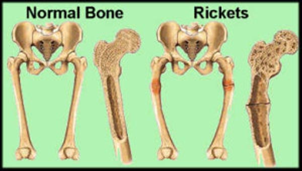
Bowed legs is not the primary indication of rickets (3 other indications of rickets are seen more often)
Rickets is typically due to low cellular Vitamin D - April 2024
Some Rickets is due to poor genes - Vitamin D needed lifelong – June 2020
Vitamin D and Rickets consensus took 80 years
Rickets category has items
Breathing
Children with pneumonia in Ethiopia were 22X more likely to have rickets – 1997
Rickets in 80 percent of infants with severe pneumonia – Sept 2010
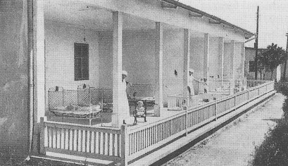
History
Majority of Medici in 1600's who died in infancy had rickets - 2013
Rickets associated with 30 percent of infant deaths in rural Netherlands in 1800’s– June 2013
Vitamin D, Cod-Liver Oil, Sunlight, and Rickets: A Historical Perspective – 2003
Vitamin D reduced so low that Victorian age diseases are returning
Turkey
400 IU vitamin D appeared to prevent infant rickets in Turkey – June 2011
Turkey gave 400 IU vitamin D to all infants and reduced Rickets by 60X - 2011
Infants again said to need more than 400 IU of vitamin D – Sept 2012 not enough in winter
Mongolia
Mongolian women have the lowest levels of vitamin D in the world – Sept 2013
Other countries
Calcium reduced some rickets in Nigerian children – Feb 2012
Vitamin D deficiency spares no body part – NE Asia – Jan 2013
Vitamin D deficiency in Middle East and North Africa - June 2013 from XML with maps
Hypovitaminosis D in the Middle East and North Africa - May 2013 from HTML
Rickets: Less costly to prevent than to treat SE Asians in UK – 2006
Rickets in 30 percent of infants in India who had low vitamin D – March 2011
Perhaps half of Russian children have rickets, 500 IU vitamin D was not enough – June 2013
Maternal vitamin D deficiency can trigger rickets in breastfed infants – review March 2013
Rickets - World
Nutritional rickets around the world: an update - 2016
- PDF is available free at Sci-Hub 10.1080/20469047.2016.1248170
- Nigeria 1 in 170; Gambia 1 in 70; Bangladesh 1 in 12
Recent Increase
Rickets in Sweden recently increased by 6X (mainly preemies) – May 2019
Rickets in Japan increased 3 X recently, similar to increases in other countries – June 2017
Rare Nutritional Rickets increased 10X in 20 years – Feb 2013
Prevention and treatment
Rickets virtually cured by 90,000 IU of Vitamin D along with daily Calcium – RCT Nov 2018
Rickets needs Vitamin D and Calcium - Global Consensus Jan 2016
Neonate Rickets was virtually always associated with very low maternal vitamin D – Dec 2014
- pehaps giving mothers Vitamin D would be better
Rickets prevented by single injection of vitamin D or weekly supplementation – RCT Jan 2014
Group is giving only 200 IU of Vitamin D to 70 million women and children to prevent Rickets
400 IU vitamin D for infants and 2000 IU during pregnancy if high risk – June 2010
Giving vitamin D to ALL children reduced (rickets) symptoms by 60 percent – Aug 2012
744 IU vitamin D needed by Northern white teens to prevent Rickets – Jan 2011
Took 4 months of 400 IU vitamin D for Iranian infants to get serum level of 30 ng – June 2013 loading dose was far faster
Rickets cured for 3 months with 10,000 IU per kg vitamin D (600,000 IU max) – Sept 2012
- Vitamin D within 3-5 days of birth prevents all rickets - Dec 2013 article at the bottom of this page
Shaken-baby syndrome
Child abuse, vitamin D deficiency, or what - for parents and defense attorneys - Cannell June 2015
Mother and father on trial for infant death – set free – death was due to rickets – Dec 2011
Lack of vitamin D in infants can result in broken bones and shaken baby syndrome - March 2010
The Vitamin Deficiency Signs That Can Send You to Prison – Feb 2014
Shaken Baby Syndrome - probably caused by Ehlers-Danlos Syndrome
Child abuse fractures – 96 percent were associated with poor bones (low vitamin D, etc.) – Oct 2019

Associated with Dark skin and breastfed
Ricket was known to be associated with dark skin and breast feeding a century ago - 2005
Black Sudanese children 350X more likely to have rickets than other Australians – April 2012
Dark Skinned babies probably need vitamin D to prevent nutritional rickets - 2001
Nutritional Rickets in Denmark especially among immigrant children- Feb 2012
16% of exclusively breastfed infants so low on vitamin D that they had rickets – June 2010
Animals
-
Rickets Other
Search VitaminDWiki for "RESISTANT RICKETS 195 items as of Dec 2018
Further look at vitamin D dependent rickets and gene mutations – Feb 2011
Vitamin D Levels in Kids are So Low that Rickets is Back with a Vengeance
A Critical Review of the Classic Metaphyseal Lesion: Traumatic or Metabolic? Jan 2014
How much Vitamin D needed to stop rickets
Rickets can be suspected below 36 ng of vitamin D – Oct 2012
Austrailia still believes 20 ng of vitamin D will provide bone health and stop rickets Oct 2013, not on VitaminDWiki
Other Vitamin D
Third study found that Infants needed 1600 IU of vitamin D – JAMA RCT May 2013
Vitamin D (40-70 ng) in Children’s Health – review Sept 2014
Breastfed infants: 90 percent had less than 20 ng of vitamin D, formula-fed: 15 percent – May 2013
Many preemies need at least 800 IU of vitamin D – RCT May 2013
75 percent of unexplained sudden infant deaths had inadequate level of vitamin D – April 2013
Overview UV and vitamin D Sun is better than UV, which is better than supplements
43 reasons for Vitamin D deficiency 19 new reasons (40 total as of 2023)
Quick, FREE, self-test for Vitamin D deficiency sore bones is one indication of rickets
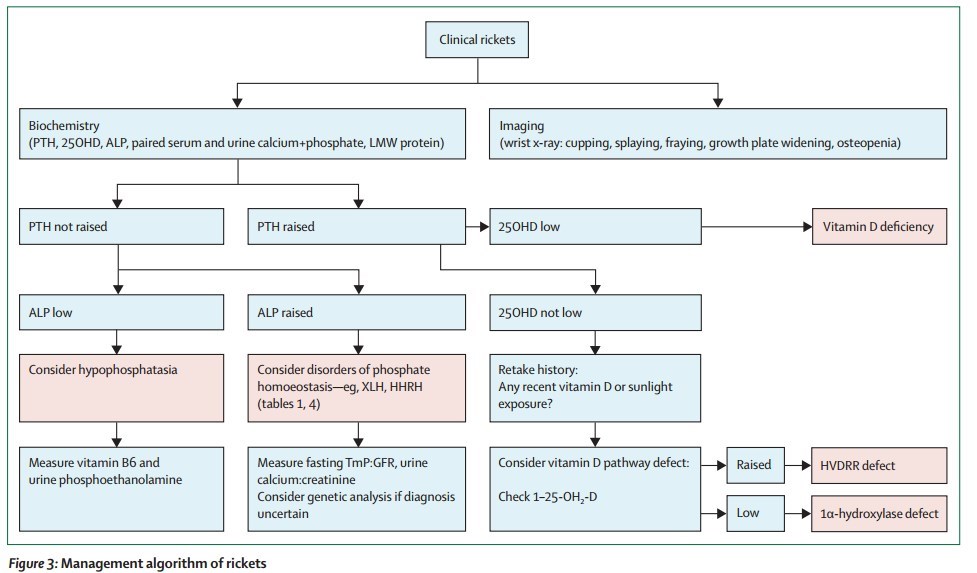
 **PDF Good Seminar on Rickets - Lancet May 2014 is attached at the bottom of this page**
**Need 400 IU**
**PDF Good Seminar on Rickets - Lancet May 2014 is attached at the bottom of this page**
**Need 400 IU**

Rickets or abuse? A histologic comparison of rickets and child abuse-related fractures
Forensic Science, Medicine, and Pathology, January 2015, $40 for PDF **Results** In the case of rickets, there was marked architectural disorganization of endochondral ossification at the costochondral junctions and growth plates of long bones. The child abuse-related fractures showed osteochondral callus at different stages of healing, either centered on a discrete fracture line or at metaphyses (e.g. classical metaphyseal lesions). In many instances, the healing fractures disrupted the line of endochondral ossification. In none of the child abuse-related fractures was there any similarity to the histologic appearance of rickets. **Conclusion** The maturation disturbance in the growth plate that occurs in rickets is a distinctive entity that cannot be confused histologically with healing fractures, including the classical metaphyseal lesion. Includes 20 references - freeVitamin D Status in Abused and Nonabused Children Younger Than 2 Years Old With Fractures - 2011
**📄 Download the PDF from VitaminDWiki**See also web
Rickets Vitamin D Council updated Aug 2014 - with PDF
Hereditary Rickets. How Genetic Alterations Explain the Biochemical and Clinical Phenotypes. Dec 2013
- hereditary rickets may be subdivided into two main groups vitamin D and phosphorus
Vitamin D receptor gene polymorphisms and the risk of rickets among Asians: a meta-analysis Jan? 2014
Disorders in Vitamin D Action Jan 2014 - mainly Rickets
Rickets is on the rise GrassrootsHealth June 2015 - nice summary
Rickets malady Malacards - has the following chart
Top 20 diseases related to Rickets by genes (many of which are associated with low vitamin D


- Unsure how valid the above 2003 diagram is in 2017
- Rickets Dec 2017 rent pdf for $5, doi:10.1038/nrdp.2017.101
- "prematurely born infants or breastfed infants who have dark skin types are particularly at risk"
- 📄 Download the Rickets Lancet 2003, PDF from VitaminDWiki
- Osteomalacia Wikipedia
- "Osteomalacia in children is known as rickets , and because of this, use of the term "osteomalacia" is often restricted to the milder, adult form of the disease"
- Nutritional rickets around the world: causes and future directions - 2006
- The roles of vitamin D and dietary calcium in nutritional rickets - 2018
In some regions late-onset Rickets may be due to low Calcium
- Nutritional rickets: deficiency of vitamin D, calcium, or both?- 2004
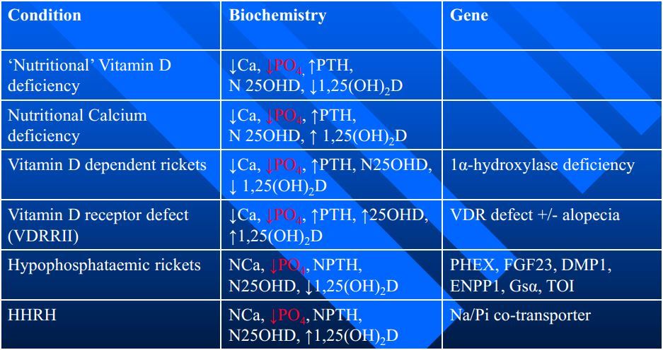
6 Types of rickets"low vitamin D, poor vitamin D receptor. etc.
Poor vitamin D Receptor limits vitamin D in blood from getting to the bones
📄 Download the PDF from VitaminDWiki
The RESISTANCE of Vitamin D Resistent Rickets may be due to Vitamin D RECEPTOR - March 2019
Detecting reasons for recurrent deformity in treatment of patients with vitamin D-resistant rickets using diagnostic imaging
J Orthop. 2019 Mar 22;16(4):325-328. doi: 10.1016/j.jor.2019.02.033. eCollection 2019 Jul-Aug.
Diachkova GV1, Novikov KI2, Effatparvar MR3, Chistova EA1, Diachkov KA1, Novikova OS1, Korkin AY4, Shikhaleva NG5.
PURPOSE: To assess age related manifestations of the femur and tibia in patients with vitamin D-resistant rickets (VDR) and explore causes for recurrent deformity using imaging modalities.
METHODS: Computed tomography (CT), magnetic resonance imaging (MRI) and dual energy X-ray densitometry (DEXA) were used to assess conditions of long bones of lower limbs in patients with vitamin D-resistant rickets aged from 4 years to 30 years preoperatively and after limb lengthening.
RESULTS:
Age related MRI findings showed specific structure of the femur and tibia in patients with (VDR) preoperatively and after operative treatment. Abundant irregular osteoid formed in femoral and tibial physes was shown to reveal complicated nature of bone deformity causing recurrence in patients with (VDR) at childhood. CT findings allowed us to detect early cortical injury, measure its length with forming Looser's zones, examine significant differences in density measurements of Looser's zones preoperatively and after deformity correction using transosseous osteosynthesis.
CONCLUSION:
Recurrent deformity can develop in patients with (VDR) due to progression of the disease, irregular osteoid deposited in the medial and lateral metaepiphysis, osteoid area measuring over 50% of epiphyseal cross section, insufficient regenerate mineralization, and formation of Looser's zones.