Vitamin D activates the hypothalamus (in rodents) to reduce weight and diabetes
Hypothalamic Vitamin D Improves Glucose Homeostasis and Reduces Weight
Diabetes. 2016 May 23. pii: db160309
Sisley SR1, Arble DM2, Chambers AP3, Gutierrez-Aguilar R4, He Y5, Xu Y5, Gardner D6, Moore DD7, Seeley RJ2, Sandoval DA2.
1Children's Nutrition Research Center, Department of Pediatrics, Baylor College of Medicine, Houston, TX 77030 Stephanie.sisley@bcm.edu.
2Department of Surgery, University of Michigan, Ann Arbor, MI 48105.
3Department of Incretin and Obesity Pharmacology, Novo Nordisk, 2760 MÅLØV, Denmark.
4División de Investigación, Facultad de Medicina, Universidad Nacional Autónoma de México Laboratorio de Enfermedades Metabólicas: Obesidad y Diabetes, Hospital Infantil de México "Federico Gómez", México.
5Children's Nutrition Research Center, Department of Pediatrics, Baylor College of Medicine, Houston, TX 77030.
6Diabetes Center, University of California San Francisco, San Francisco, CA 94143.
7Department of Molecular and Cellular Biology, Baylor College of Medicine, Houston, TX 77030.
Despite clear associations between vitamin D deficiency and obesity and/or type 2 diabetes, a causal relationship is not established. Vitamin D receptors (VDR) are found within multiple tissues, including the brain. Given the importance of the brain in controlling both glucose levels and body weight, we hypothesized that activation of central VDR links vitamin D to the regulation of glucose and energy homeostasis. Indeed, we found that small doses of active vitamin D, 1,25D3 (calcitriol), into the third-ventricle of the brain improved glucose tolerance and markedly increased hepatic insulin sensitivity, an effect that is dependent upon VDR within the paraventricular nucleus of the hypothalamus.
In addition, chronic central administration of 1,25D3 dramatically decreased body weight by lowering food intake in obese rodents.
Our data indicate 1,25D3-mediated changes in food intake occurs through action within the arcuate nucleus. We found that VDR co-localized with and activated key appetite regulating neurons in the arcuate, namely pro-opiomelanocortin neurons (POMC). Together, these findings define a novel pathway for vitamin D regulation of metabolism with unique and divergent roles for CNS VDR signaling. Specifically, our data suggest that vitamin D regulates glucose homeostasis via the paraventricular nuclei and energy homeostasis via the arcuate nuclei.
© 2016 by the American Diabetes Association. PMID: 27217488
Publisher wants $35 for the PDF
Comparison of thalamus and hypothalamus
Chat-GPT Oct 2027
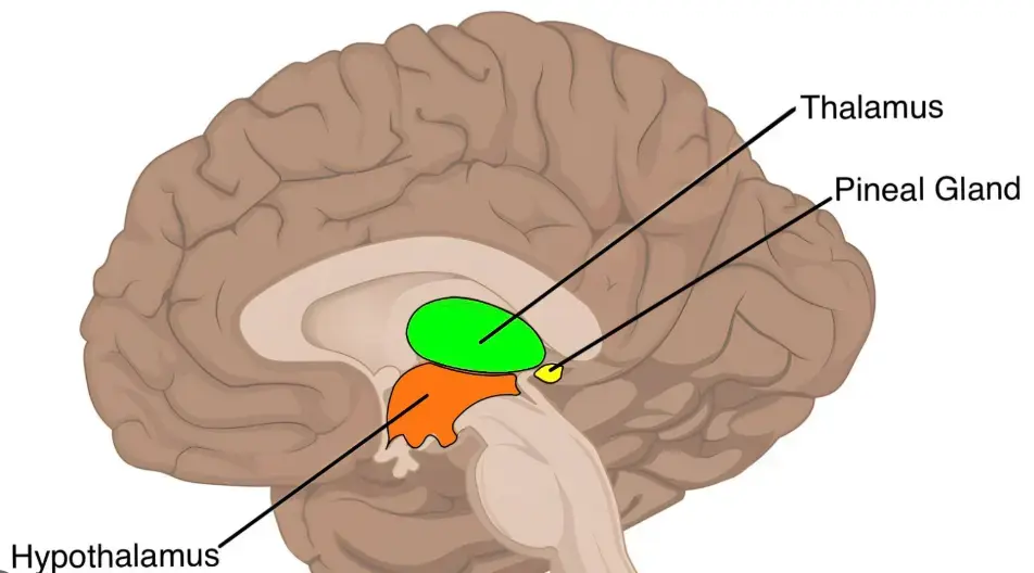
The thalamus and hypothalamus are two closely connected structures deep within the brain. Although their names are similar, their functions are quite different.
- Thalamus: Primarily processes and routes sensory and motor information and helps regulate consciousness, attention, and sleep.
- Hypothalamus: Regulates the body's internal environment, including temperature, hunger, thirst, hormones, and autonomic nervous system activity.
A useful analogy is that the thalamus is an information-routing center, while the hypothalamus is the body's automatic control center.
1. Location in the brain
23.1: Foundations of Neuromuscular Functioning - Medicine LibreTexts
Motor and Sensory Functions of the Nervous System
Both structures are part of the diencephalon, located between the cerebral hemispheres and the brainstem. The hypothalamus lies immediately below and slightly forward of the thalamus.
2. Comparison of functions
| Function | Thalamus | Hypothalamus |
|---|---|---|
| Primary role | Information processing and relay | Internal body regulation |
| Sensory information | Major role, including vision, hearing, touch, pain | Limited direct role |
| Motor control | Relays signals involving cerebellum and basal ganglia | Indirect influence |
| Body temperature | Minimal direct role | Major control center |
| Hunger and appetite | Limited | Major role |
| Thirst and water balance | Limited | Major role |
| Sleep | Sleep rhythms and arousal | Circadian timing and sleep–wake regulation |
| Hormones | Limited direct role | Controls pituitary hormone release |
| Blood pressure | Indirect influence | Autonomic regulation |
| Emotions | Participates in emotional circuits | Coordinates physical responses to emotion |
| Memory | Important circuits | Some specialized involvement |
| Consciousness | Essential for normal arousal and awareness | Influences arousal |
3. How they work
Thalamus — processing information
Most sensory signals pass through thalamic nuclei before reaching the cerebral cortex. Smell is a notable exception to this initial relay pattern. The thalamus also participates in attention, motor coordination, and maintaining consciousness.
23.1: Foundations of Neuromuscular Functioning - Medicine LibreTexts
Hypothalamus — maintaining homeostasis
Monitors internal conditions and coordinates responses through the autonomic nervous system and pituitary gland. It helps regulate temperature, metabolism, fluid balance, stress responses, and reproductive hormones.
4. What happens when something goes wrong?
| Symptom or disorder | Thalamus involvement | Hypothalamus involvement |
|---|---|---|
| Dizziness or vertigo | Possible, especially with certain lesions | Possible, often indirectly |
| Migraine | Involved in sensory and pain processing | Involved in migraine initiation and associated symptoms |
| Nausea and vomiting | Indirect role | Autonomic involvement |
| Sleep disorders | Disrupted sleep rhythms | Disrupted circadian timing |
| Chronic pain | Central post-stroke pain and sensory dysfunction | Modulation of pain and stress |
| Temperature instability | Uncommon | Characteristic |
| Hormonal abnormalities | Uncommon | Characteristic |
| Excessive thirst or urination | Uncommon | Possible with ADH regulation problems |
| Memory difficulties | Possible | Possible in certain lesions |
5. Particularly interesting: their roles in migraine
Both structures are involved in migraine, but in different ways.
Hypothalamus
Research suggests that hypothalamic activity can change before migraine pain begins. This may help explain early symptoms such as yawning, fatigue, thirst, food cravings, nausea, and changes in sleep.
Thalamus
The thalamus participates in processing migraine-related pain and sensory hypersensitivity. It helps explain why some people with migraines become unusually sensitive to light, sound, or touch.
The two structures also communicate with brainstem networks that participate in nausea, vomiting, vestibular processing, and autonomic responses.
An important distinction is that neither structure alone explains all migraine symptoms. Vestibular migraine, for example, involves broader networks including the vestibular nuclei, cerebellum, thalamus, and cortex.
6. Relative size
The thalamus is substantially larger than the hypothalamus.
- Thalamus: Two approximately egg-shaped masses, one on each side of the third ventricle, each roughly 3–4 cm long.
- Hypothalamus: A much smaller collection of nuclei beneath the thalamus, weighing only a few grams in total.
Despite its small size, the hypothalamus has enormous influence over the body's physiological functions.
One especially interesting question is how the hypothalamus and thalamus interact during severe migraine episodes involving vertigo, nausea, and vomiting. The hypothalamus may contribute to the autonomic and premonitory symptoms, while the thalamus helps integrate vestibular and sensory signals.