Poor CYP genes affect Vitamin D and several other systems in the body
Rare Mendelian CYP Disorders: From Metabolic Pathways to Biomarker-Guided Diagnosis
Drug Metab Rev . 2026 Oct 12:1-37. doi: 10.1080/03602532.2026.2743901. PDF behind paywall
Wojciech Kuban 1, Ewa Bromek 1, Renata Pukło 1, Anna Haduch 1
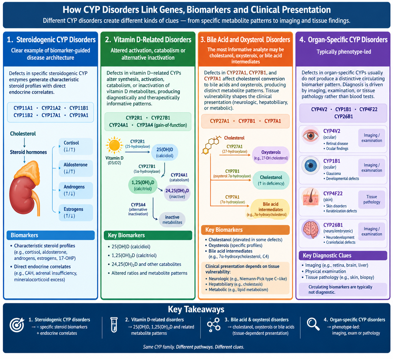
Cytochrome P450 (CYP) enzymes are best known for xenobiotic metabolism, but many human CYPs also catalyze essential reactions in endogenous pathways governing steroidogenesis, bile acid and oxysterol turnover, vitamin D homeostasis, retinoic acid degradation, and tissue-specific lipid metabolism. Pathogenic variation in these enzymes gives rise to a distinctive class of rare Mendelian disorders in which enzyme position within a pathway predicts the dominant biochemical or tissue-restricted readout and, consequently, the clinical phenotype. Here, we synthesize rare CYP-related diseases within a pathway-resolved framework and propose that they are best interpreted not as isolated syndromes, but as tightly coupled metabolic systems linking gene defects, enzymatic block, metabolite signature, and disease expression.
Steroidogenic CYP disorders provide the clearest example of biomarker-guided disease architecture, as defects in CYP11A1, CYP21A2, CYP11B1, CYP11B2, CYP17A1, and CYP19A1 generate characteristic steroid profiles with direct endocrine correlates. Vitamin D-related disorders involving CYP2R1, CYP27B1, CYP24A1, and gain-of-function CYP3A4 variants illustrate how altered activation, catabolism, or alternative inactivation produces diagnostically and therapeutically informative metabolite patterns. Bile acid and oxysterol disorders caused by CYP27A1, CYP7B1, and CYP7A1 defects show that the most informative analyte may be cholestanol, oxysterols, or bile acid intermediates, whereas tissue vulnerability shapes neurologic, hepatobiliary, or metabolic presentation. By contrast, organ-specific CYP disorders involving CYP4V2, CYP1B1, CYP4F22, and CYP26B1 are typically phenotype-led, with diagnosis driven by imaging, examination, or tissue pathology rather than circulating biomarkers.
(Bolded text was given to 3 AIs)
Across categories, mechanistic resolution depends on aligning pathway architecture with the appropriate biochemical readout and locus-aware molecular interrogation.
IClaude AI - Opus
CYP Disorders: Biomarker-Led to Phenotype-Led Diagnosis
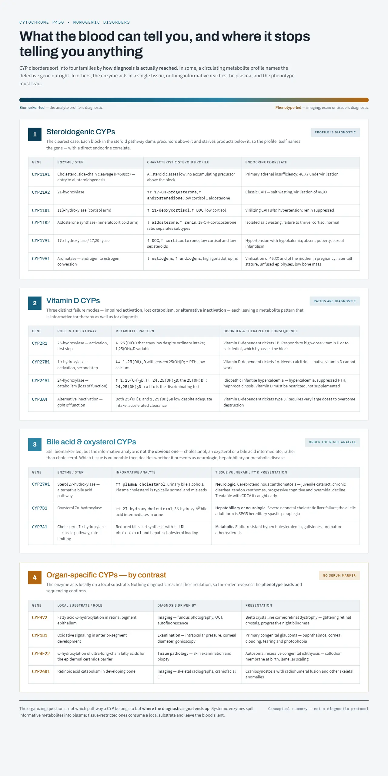
CYP disorders sort into four families by how diagnosis is actually reached. Systemic enzymes spill informative metabolites into plasma, so the analyte profile names the gene. Tissue-restricted enzymes consume a local substrate and leave the blood silent, so the phenotype must lead and sequencing confirms.
Overview
| Group | Genes | Diagnosis driven by | Where the signal is |
|---|---|---|---|
| Steroidogenic | CYP11A1; CYP21A2; CYP11B1; CYP11B2; CYP17A1; CYP19A1 | Circulating steroid profile | Plasma — profile names the gene |
| Vitamin D | CYP2R1; CYP27B1; CYP24A1; CYP3A4 | Metabolite levels and ratios | Plasma — pattern guides therapy too |
| Bile acid and oxysterol | CYP27A1; CYP7B1; CYP7A1 | A specific sterol intermediate | Plasma — but not the obvious analyte |
| Organ-specific | CYP4V2; CYP1B1; CYP4F22; CYP26B1 | Imaging, examination, tissue pathology | Tissue — no circulating marker |
1. Steroidogenic CYPs
Each block in the steroid pathway dams precursors above it and starves products below it, so the profile itself names the gene, with a direct endocrine correlate.
| Gene | Enzyme / step | Characteristic steroid profile | Endocrine correlate |
|---|---|---|---|
| CYP11A1 | Cholesterol side-chain cleavage (P450scc); entry to all steroidogenesis | All steroid classes low; no precursor accumulates above the block | Primary adrenal insufficiency; 46,XY undervirilization |
| CYP21A2 | 21-hydroxylase | ↑↑ 17-OH-progesterone; ↑ androstenedione; low cortisol ± aldosterone | Classic CAH; salt wasting; virilization of 46,XX |
| CYP11B1 | 11β-hydroxylase; cortisol arm | ↑ 11-deoxycortisol; ↑ DOC; low cortisol | Virilizing CAH with hypertension; renin suppressed |
| CYP11B2 | Aldosterone synthase; mineralocorticoid arm | ↓ aldosterone; ↑ renin; 18-OH-corticosterone ratio separates subtypes | Isolated salt wasting; failure to thrive; cortisol normal |
| CYP17A1 | 17α-hydroxylase / 17,20-lyase | ↑ DOC; ↑ corticosterone; low cortisol and low sex steroids | Hypertension with hypokalemia; absent puberty; sexual infantilism |
| CYP19A1 | Aromatase; androgen to estrogen conversion | ↓ estrogens; ↑ androgens; ↑ gonadotropins | 46,XX virilization and maternal virilization in pregnancy; later tall stature, unfused epiphyses, low bone mass |
2. Vitamin D CYPs
Three distinct failure modes — impaired activation, lost catabolism, or alternative inactivation — each leaving a metabolite pattern informative for therapy as well as diagnosis.
| Gene | Role in the pathway | Metabolite pattern | Disorder and therapeutic consequence |
|---|---|---|---|
| CYP2R1 | 25-hydroxylase; activation step 1 | ↓ 25(OH)D that stays low despite ordinary intake; 1,25(OH)₂D variable | Vitamin D-dependent rickets 1B; responds to high-dose vitamin D or to calcifediol, which bypasses the block |
| CYP27B1 | 1α-hydroxylase; activation step 2 | ↓↓ 1,25(OH)₂D with normal 25(OH)D; ↑ PTH; low calcium | Vitamin D-dependent rickets 1A; needs calcitriol, since native vitamin D cannot work |
| CYP24A1 | 24-hydroxylase; catabolism, loss of function | ↑ 1,25(OH)₂D; ↓↓ 24,25(OH)₂D; high 25(OH)D to 24,25(OH)₂D ratio is the discriminating test | Idiopathic infantile hypercalcemia; suppressed PTH, nephrocalcinosis; vitamin D must be restricted, not supplemented |
| CYP3A4 | Alternative inactivation of 25(OH)D and 1,25(OH)₂D; gain of function | Both metabolites low despite adequate intake; accelerated clearance | Vitamin D-dependent rickets type 3; requires very large doses to overcome destruction |
3. Bile Acid and Oxysterol CYPs
Still biomarker-led, but the informative analyte is not the obvious one — cholestanol, an oxysterol or a bile acid intermediate rather than cholesterol. Tissue vulnerability then decides whether presentation is neurologic, hepatobiliary or metabolic.
| Gene | Enzyme / step | Informative analyte | Tissue vulnerability and presentation |
|---|---|---|---|
| CYP27A1 | Sterol 27-hydroxylase; alternative bile acid pathway | ↑↑ plasma cholestanol; urinary bile alcohols; plasma cholesterol typically normal and misleading | Neurologic — cerebrotendinous xanthomatosis: juvenile cataract, chronic diarrhea, tendon xanthomas, progressive cognitive and pyramidal decline; treatable with CDCA if caught early |
| CYP7B1 | Oxysterol 7α-hydroxylase | ↑↑ 27-hydroxycholesterol; 3β-hydroxy-Δ⁵ bile acid intermediates in urine | Hepatobiliary or neurologic — severe neonatal cholestatic liver failure; the allelic adult form is SPG5 hereditary spastic paraplegia |
| CYP7A1 | Cholesterol 7α-hydroxylase; classic pathway, rate-limiting | Reduced bile acid synthesis; ↑ LDL cholesterol; hepatic cholesterol loading | Metabolic — statin-resistant hypercholesterolemia, gallstones, premature atherosclerosis |
4. Organ-Specific CYPs
By contrast: the enzyme acts locally on a local substrate, nothing diagnostic reaches the circulation, and the order reverses — phenotype leads, sequencing confirms.
| Gene | Local substrate / role | Diagnosis driven by | Presentation |
|---|---|---|---|
| CYP4V2 | Fatty acid ω-hydroxylation in retinal pigment epithelium | Imaging — fundus photography, OCT, autofluorescence | Bietti crystalline corneoretinal dystrophy; glittering retinal crystals, progressive night blindness |
| CYP1B1 | Oxidative signaling in anterior-segment development | Examination — intraocular pressure, corneal diameter, gonioscopy | Primary congenital glaucoma; buphthalmos, corneal clouding, tearing and photophobia |
| CYP4F22 | ω-hydroxylation of ultra-long-chain fatty acids for the epidermal ceramide barrier | Tissue pathology — skin examination and biopsy | Autosomal recessive congenital ichthyosis; collodion membrane at birth, lamellar scaling |
| CYP26B1 | Retinoic acid catabolism in developing bone | Imaging — skeletal radiographs, craniofacial CT | Craniosynostosis with radiohumeral fusion and other skeletal anomalies |
Organizing Principle
The question is not which pathway a CYP belongs to, but where the diagnostic signal ends up. Groups 1 to 3 are biomarker-led and differ only in how obvious the right analyte is. Group 4 is phenotype-led because the enzyme never releases an informative metabolite into the blood.
Conceptual summary — not a diagnostic protocol.
Gemini
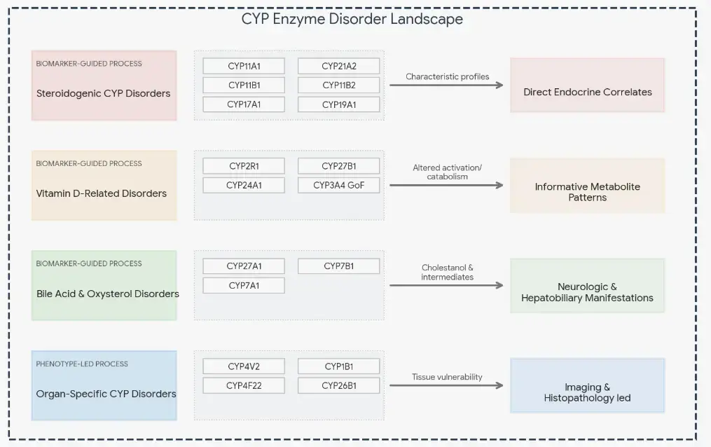
This infographic architecture breaks down the clinical and biochemical relationships of Cytochrome P450 (CYP) disorders into two distinct paradigms: biomarker-guided systems (where circulating molecules drive diagnosis) and phenotype-led systems (where structural tissue damage drives diagnosis).
The Two Diagnostic Paradigms of CYP Disorders
1. Biomarker-Guided Disease Architecture
In these disorders, specific genetic defects yield predictable biochemical signatures in blood or urine, acting as a direct map to clinical presentation.
- Steroidogenic CYP Disorders
- Enzymes Involved: CYP11A1, CYP21A2, CYP11B1, CYP11B2, CYP17A1, CYP19A1
- Mechanism: Defects block specific pathways in steroidogenesis, causing upstream accumulation and downstream deficiencies.
- Clinical Correlate: Yields characteristic steroid profiles (e.g., elevated 17-hydroxyprogesterone in 21-hydroxylase deficiency) with direct endocrine outcomes like adrenal insufficiency or virilization.
- Vitamin D-Related Disorders
- Enzymes Involved: CYP2R1, CYP27B1, CYP24A1, and gain-of-function (GoF) CYP3A4 variants
- Mechanism: Disrupts the tight equilibrium of homeostatic activation (hydroxylation) and catabolic elimination.
- Clinical Correlate: Generates therapeutically actionable metabolite patterns, differentiating between impaired activation (rickets) and defective catabolism (idiopathic infantile hypercalcemia).
- Bile Acid & Oxysterol Disorders
- Enzymes Involved: CYP27A1, CYP7B1, CYP7A1
- Mechanism: Blocks cholesterol clearance and bile acid synthesis pathways, accumulating toxic intermediates.
- Clinical Correlate: Diagnosis relies on measuring unique analytes like cholestanol or plasma oxysterols, while differential tissue vulnerability dictates whether a patient presents with infant cholestasis or adult-onset neurodegeneration (e.g., Cerebrotendinous Xanthomatosis).
2. Phenotype-Led Architecture
Unlike systemic metabolic pathways, these localized enzymes do not typically leave a recognizable chemical footprint in systemic circulation.
- Organ-Specific CYP Disorders
- Enzymes Involved: CYP4V2 (Bietti crystalline dystrophy), CYP1B1 (congenital glaucoma), CYP4F22 (lamellar ichthyosis), CYP26B1 (skeletal anomalies)
- Mechanism: Localized metabolic failure causes microenvironment disruption or structural cellular toxicity.
- Clinical Correlate: Diagnosis cannot rely on plasma biomarkers. It is entirely phenotype-led, requiring clinical examinations, advanced imaging (e.g., optical coherence tomography), or tissue pathology.
Chat-GPT