
Ectopic pregnancies: 5X increase in 50 years (now 2%), 6.4 X more likely if Vitamin D < 30 ng
What is an ectopic pregnancy
Claude AI Sept 2026
An ectopic pregnancy is one where the fertilized ovum implants outside the uterine cavity. About 98% are tubal — most often in the ampulla of the fallopian tube — with the rest in the interstitial/cornual segment, ovary, cervix, abdomen, or a cesarean scar. None are viable, and the danger is rupture with intraperitoneal hemorrhage, which is still a leading cause of first-trimester maternal death.
The underlying mechanism is usually a transport failure: damaged tubal cilia or altered smooth-muscle activity delays the embryo's passage, and it implants where it stalls. That is why the dominant risk factors are things that scar or inflame the tube — prior pelvic inflammatory disease (chlamydia especially), prior tubal surgery, endometriosis, smoking (which impairs ciliary beat), and ART/IVF. A previous ectopic raises risk roughly 10–15 fold and PID 6–10 fold.
Incidence: 2% now, had been 0,4%
Roughly 2% of reported pregnancies in the US, about 1 in 50 worldwide, with developing-country rates as high as 27.7 per 1,000 deliveries. A large Kaiser Permanente Northern California analysis put it at 1.53% (95% CI 1.40–1.67). Older figures from developed countries give 11–20 per 1,000 live births.
The trend is interesting: incidence rose from about_ 0.4% to 2% over the past 50 years_ in developed nations — largely attributed to better detection (transvaginal ultrasound plus sensitive β-hCG), more chlamydia, and more IVF. Globally, though, the age-standardized rate has been falling: from 131.9 per 100,000 in 1990 to 84.3 per 100,000 in 2019, with the burden concentrated in low-SDI regions.
Vitamin D association with ectopic pregnancies
Thin but consistent — two case-control studies, both from Iran, and nothing else of substance that I can find.
Sahhaf et al. 2019 (150 ectopic vs. 150 normal pregnancies): vitamin D was significantly higher in controls (p = 0.002); across all 300 patients, 60.7% were deficient and 21.3% insufficient. [Sahhaf 2019]
Sarani et al. 2022 (51 vs. 51, first trimester): controls averaged 34.31 ± 7.32 ng/mL vs. 20.95 ± 20.68 ng/mL in the ectopic group (p < 0.001); women under 30 ng/mL had 6.40× the odds of ectopic pregnancy (95% CI 3.26–15.83). Age, BMI, and parity did not differ between groups. [Sarani 2022]
Two notes
- An SD of 20.68 on a mean of 20.95 is implausible for a normally-distributed 25(OH)D measurement in 51 women, and the reported CI (3.26–15.83) isn't geometrically centered on 6.40 the way a log-scale OR interval should be. The direction of effect is probably real; the magnitude I'd treat as unreliable.
- No RCT, no cohort, no Mendelian randomization on this endpoint — I checked. So the causal question is open.
Why it's biologically plausible anyway. Ectopic pregnancy is largely a tubal-damage disease, and the main cause of tubal damage is chlamydial infection and the inflammatory response to it. Vitamin D's best-established non-skeletal role is innate immunity and modulation of the inflammatory response — cathelicidin induction, damping of the Th1/Th17 response that drives scarring. If low D increases susceptibility to or severity of PID, ectopic risk follows downstream without vitamin D acting on the tube at all. There's also direct expression of VDR and CYP27B1 in fallopian tube epithelium and decidua, which would allow a local effect on implantation timing, but that's speculative here.
Confounding to flag: low 25(OH)D travels with smoking, obesity, low SES, and higher STI exposure — all independent ectopic risk factors. A case-control design can't separate those, and neither Iranian study adjusted for smoking or infection history.
Probably need >30 ng of vitamin D at time of conception
One practical implication worth noting: unlike preeclampsia or gestational diabetes, ectopic risk is set before and at implantation. Supplementation started at the first prenatal visit is already too late by definition — the exposure window that matters is preconception, which is exactly what the Sarani authors concluded.
Ectopic Pregnancy and Vitamin D studies
- Association of serum vitamin D levels with ectopic pregnancy - Jan 2023 PDF 6.4 X more likely if Vitamin D < 30 ng
- Relationship Between Serum Vitamin D Level and Ectopic Pregnancy Sept 2019 PDF
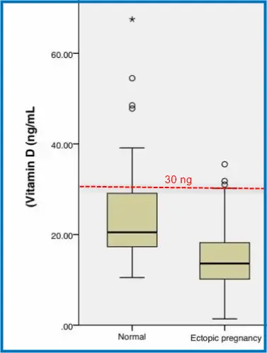
Risk of Ectopic Pregnancies increased with Vitamin D - Sept 2026 (disagrees with others)
The causal associations between 25(OH) vitamin D levels and ectopic pregnancy: A 2-sample Mendelian randomization study
Medicine (Baltimore). 2026 Sep 18;105(38) doi: 10.1097/MD.0000000000050657 PDF
Pei-Jin Zhang 1, Shao Bo 2, Shuang Liang 1, Hong-Yuan Zhang 1, Xiao-Dong Fan 1 Affiliations Expand
Previous research has indicated a potential link between 25-hydroxyvitamin D [25(OH)D] and ectopic pregnancy (EP), but the underlying causal relationship remains unclear. We performed a 2-sample Mendelian randomization (MR) study to investigate the causal association between circulating 25(OH)D and ectopic pregnancy (EP). The primary analysis used summary statistics from a 25(OH)D GWAS of 4,96,946 European-ancestry participants and FinnGen summary statistics for EP (3111 cases and 89,340 controls). To assess whether the finding was robust to the choice of 25(OH)D GWAS, we repeated the MR analysis using a separate 25(OH)D GWAS (n = 4,41,291 European-ancestry participants) as a replication analysis; it was not used solely as a within-analysis sensitivity test. The primary and replication analyses used inverse-variance weighting (IVW) as the main method, complemented by weighted mode, simple mode, weighted median, and MR-Egger analyses. Heterogeneity and pleiotropy assessments, leave-one-out analyses, and other sensitivity analyses were conducted within each MR analysis. IVW estimates showed that increased serum 25(OH)D levels promoted EP (OR = 1.323, 95% CI = 1.024-1.710, P = .032). The replication analysis also revealed similar trends (OR = 1.383, 95% CI = 1.041-1.836, P = .025). The primary and replicated analyses were combined in a meta-analysis, and the result showed a significant causal influence (OR = 1.350, 95% CI = 1.116-1.632, P = .002). Sensitivity analysis confirmed the robustness of the MR estimations. In this MR study of European-ancestry participants, genetically predicted higher circulating 25(OH)D levels were associated with a higher risk of EP. These findings do not establish serum 25(OH)D as a diagnostic marker for EP. Rather, if confirmed in further clinical studies, elevated vitamin D may be considered a potential associated risk factor only in the context of established clinical signs and symptoms of EP.
Related in VitaminDWiki
- Ensure a healthy pregnancy and baby - take Vitamin D before conception
Ectopic pregnancies are probably like a miscarriage - need Vitamin D very early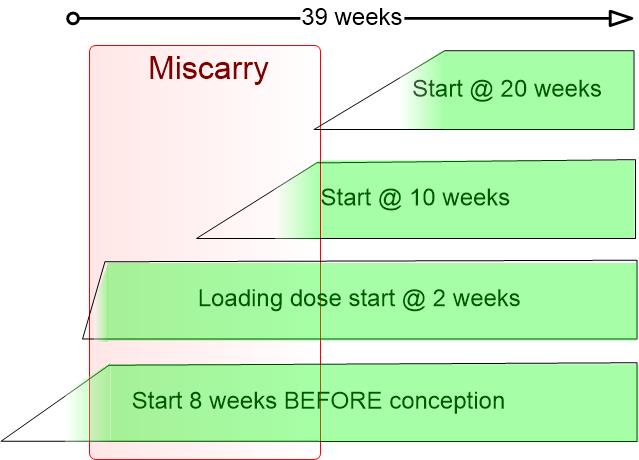
- Miscarriage and low Vitamin D – many studies
- Recurrent pregnancy loss (miscarriage) is associated with low vitamin D in 6 ways
Comparison of Ectopic Pregnancies and Miscarriages
Claude AI Sept 2026
Ectopic pregnancy vs miscarriage — a side-by-side comparison
Both end a pregnancy in the first trimester, and low vitamin D has been associated with both. But they differ in almost every other respect that matters — how often they happen, whether the mother is in danger, and how good the vitamin D evidence actually is. The evidence quality is asymmetric enough that it is flagged as its own row rather than buried in a footnote.
Comparison table
| Ectopic pregnancy | Miscarriage (spontaneous abortion) | |
|---|---|---|
| Definition | Implantation outside the uterine cavity — about 98% tubal, mostly in the ampulla; the rest interstitial, ovarian, cervical, abdominal, or in a cesarean scar. | Loss of an intrauterine pregnancy before 20 weeks. The first-trimester form is a nonviable intrauterine pregnancy with an empty sac or absent fetal heart activity through 12 6/7 weeks. |
| Incidence | About 2% of reported US pregnancies, roughly 1 in 50 worldwide. Kaiser Permanente Northern California: 1.53% (95% CI 1.40–1.67). Developing-country rates reach 27.7 per 1,000 deliveries. | About 10% of clinically recognized pregnancies (ACOG); 15–20% by most estimates; 30–60% of all conceptions once unrecognized biochemical losses are counted. |
| 50-year trend | Rose roughly 5×, from about 0.4% to 2% in developed nations. Globally the age-standardized rate is falling: 131.9 per 100,000 in 1990 to 84.3 in 2019, with the burden concentrated in low-SDI regions. | Essentially flat. No comparable secular rise has been reported. |
| Time after conception | Nearly always first trimester. Diagnosis typically 3–7 weeks after conception (5–9 weeks from LMP); rupture usually by about 8 weeks after conception. | About 80% of losses occur in the first trimester; only 1–5% fall between 12 and 20 weeks. The peak is 4–8 weeks after conception. |
| Risk of mother's death | The dangerous one. US mortality about 0.5 deaths per 100,000 live births; 3–4% of all pregnancy-related deaths; the leading cause of first-trimester pregnancy-related death. Case-fatality fell from about 35 to 3.4 per 10,000 ectopics between 1970 and 1987. African hospital series report 1–3%, roughly 10× the developed-world rate. In a Michigan death review, 44% of the women who died were found dead at home or dead on arrival. | Very low wherever care is available. Deaths arise from hemorrhage or sepsis in incomplete or septic loss, not from the loss itself. |
| Risk of fetal death | Effectively 100%. No ectopic pregnancy is viable. | 100% by definition — the loss is the outcome. |
| Increased risk if low vitamin D | Sarani 2022 (51 vs 51): controls averaged 34.31 ± 7.32 ng/mL against 20.95 ± 20.68 ng/mL in the ectopic group, p < 0.001; odds ratio 6.40 (95% CI 3.26–15.83) below 30 ng/mL, with age, BMI and parity not differing between groups. Sahhaf 2019 (150 vs 150): controls higher at p = 0.002, but no group means and no effect size reported. Bahat 2020 (50 vs 53, Turkey): 11.09 vs 14.75 ng/mL, p = 0.006, with calcium and magnesium also lower. | Deficient (<20 ng/mL) vs replete (>30 ng/mL): odds ratio 1.94 (1.25–3.02), 4 studies, n = 3,674. Insufficient and deficient combined: 1.60 (1.11–2.30), 6 studies, n = 6,338. Recurrent loss: 4.02 (2.23–7.25), 14 studies. |
| Strength of that evidence | Three case-control studies, about 500 women in total, all from populations where most women are deficient regardless of outcome (Iran twice, Turkey once). No cohort, no RCT, no Mendelian randomization. None adjusted for smoking or infection history. | Two meta-analyses agreeing in direction, but graded "low" or "very low" under GRADE, and a Mendelian randomization study found no causal association (OR 0.995, p = 0.93). |
| Exposure window | Preconception and implantation. Risk is fixed before a first prenatal visit can happen. | Preconception through early first trimester. Most losses precede the 8-week visit. |
| Proposed mechanism | Tubal transport failure. VDR, CYP27B1, CYP24A1, RXR and the calcium-sensing receptor are expressed in fallopian tube and are significantly altered at the implantation site. A plausible indirect route runs low vitamin D to worse chlamydial PID to tubal scarring, with no local action on the tube required. | Immune regulation at the maternal-fetal interface, decidualization, and placental 1,25(OH)2D signaling. |
| Other risk factors | Prior ectopic (10–15×), PID (6–10×), prior tubal surgery, endometriosis, smoking, ART. Risk is 3–4× higher at ages 35–44 than at 15–24. | Chromosomal abnormality causes 80–90% of first-trimester losses; then maternal age, obesity, smoking. |
| Recurrence | About 10–15% after one ectopic. | One loss in about 10.8% of women, two in 1.9%, three or more in 0.7%. |
Reading the vitamin D evidence honestly
The largest effect size comes from the weakest study
The ectopic odds ratio of 6.40 below 30 ng/mL is the biggest number on this page. It is three times the miscarriage figure of 1.94 and larger even than recurrent loss at 4.02. That ordering should increase suspicion rather than confidence. The 6.40 rests on 51 cases; the 1.94 pools 3,674 women. Small case-control studies drawn from high-deficiency populations systematically produce the largest effect sizes, because reverse causation and unmeasured confounding push in the same direction and there is no sample size to average them out.
Two specific problems with the Sarani numbers are worth stating plainly rather than leaving for a critic to find:
- A standard deviation of 20.68 on a mean of 20.95 ng/mL is not plausible for a normally distributed 25(OH)D measurement in 51 women.
- The confidence interval 3.26–15.83 is not geometrically centered on 6.40, which a log-scale odds ratio interval should be.
The direction of effect is probably real. The magnitude should be treated as unreliable.
What the Sahhaf percentages actually describe
Sahhaf 2019 reports that 60.66% of patients were deficient and 21.33% insufficient — about 82% below 30 ng/mL. That figure covers all 300 women, cases and controls together. It describes vitamin D status in Tabriz, not ectopic risk, and it should not be quoted as a property of the ectopic group. The study's usable contribution is the direction of difference at p = 0.002; it published no group means and no odds ratio, so no effect size can be extracted from it.
The 5× rise cuts both ways
Vitamin D status in developed nations did decline over the same fifty years that ectopic incidence rose from 0.4% to 2%, so the correlation is real. But miscarriage rates stayed flat across that identical window. If low vitamin D drove implantation failure, both curves would be expected to move.
The conventional explanation covers the ectopic rise without invoking vitamin D at all: before transvaginal ultrasound and sensitive beta-hCG assays, most early ectopics resolved undiagnosed as a late heavy period, exactly as most early miscarriages still do. Rising chlamydia rates and IVF add real cases on top of the detection effect. The 5× figure is best presented as context for why the condition matters now, not as evidence of a vitamin D effect — the flat miscarriage line is available to anyone who wants to argue the other way.
The argument that survives
The preconception timing argument does not depend on whether the odds ratio is 6.40 or 1.60. Ectopic risk is fixed at implantation, roughly two weeks after conception and typically weeks before a woman knows she is pregnant. Supplementation begun at a first prenatal visit cannot reach that window. That is a structural argument from timing rather than from a contested effect size, and it holds even if Sarani's magnitude is wrong by a factor of three.
References
- Sarani et al., Association of serum vitamin D levels with ectopic pregnancy, J Family Med Prim Care 2022 [Sarani 2022]
- Sahhaf et al., Relationship between serum vitamin D level and ectopic pregnancy, J Family Reprod Health 2019 [Sahhaf 2019]
- Bahat et al., The role of serum vitamin D and calcium levels in ectopic pregnancies, GREM 2020 [Bahat 2020]
- Refaat et al., Vitamin D-related molecules and calcium-sensing receptor in human fallopian tube, Cell Tissue Res 2017 [Refaat 2017]
- Tamblyn et al., Vitamin D and miscarriage: a systematic review and meta-analysis, Fertil Steril 2022 [Tamblyn meta]
- Chen et al., Serum vitamin D and recurrent spontaneous abortion: meta-analysis 2022 [Chen RSA meta]
- Zhang et al., No causal relationship between miscarriage and 25(OH)D: a Mendelian randomization study, Hum Reprod Open 2024 [MR study]
- ACOG Practice Bulletin, Early Pregnancy Loss [ACOG]
- Prevalence of sporadic and recurrent pregnancy loss, Fertil Steril [Loss prevalence]
- Ectopic pregnancy epidemiology overview [Medscape ectopic]
- Creanga et al., Trends in ectopic pregnancy mortality in the United States 1980-2007 [EP mortality trend]
- CDC MMWR, Ectopic pregnancy case-fatality, United States 1987 [CDC 1987]
- Ectopic pregnancy incidence, Kaiser Permanente Northern California [Permanente J]
- Global burden of ectopic pregnancy 1990-2019 [Global burden]
Compiled September 2026.