2260 diseases found to be fought by fully-activated Vitamin D (Calcitriol)
Effects of vitamin D signaling in cardiovascular disease: centrality of macrophage polarization
Front Cardiovasc Med. 2024 Jun 25:11:1388025. doi: 10.3389/fcvm.2024.1388025
Anton Fliri 1, Shama Kajiji 1 founders of Emergent System Analytics
"Unlocking the power of proteomics, emergent illuminates a personalized path to health, where precision meets potential, and every individual's unique biology becomes the cornerstone of their well-being."
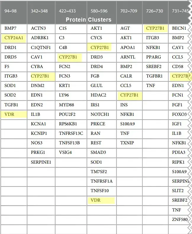
A small portion of the proteins affected by Calcitriol
Among the leading causes of natural death are cardiovascular diseases, cancer, and respiratory diseases. Factors causing illness include genetic predisposition, aging, stress, chronic inflammation, environmental factors, declining autophagy, and endocrine abnormalities including insufficient vitamin D levels. Inconclusive clinical outcomes of vitamin D supplements in cardiovascular diseases demonstrate the need to identify cause-effect relationships without bias. We employed a spectral clustering methodology capable of analyzing large diverse datasets for examining the role of vitamin D's genomic and non-genomic signaling in disease in this study. The results of this investigation showed the following:
(1) vitamin D regulates multiple reciprocal feedback loops including p53, macrophage autophagy, nitric oxide, and redox-signaling;
(2) these regulatory schemes are involved in over 2,000 diseases.
Furthermore, the balance between genomic and non-genomic signaling by vitamin D affects autophagy regulation of macrophage polarization in tissue homeostasis. These findings provide a deeper understanding of how interactions between genomic and non-genomic signaling affect vitamin D pharmacology and offer opportunities for increasing the efficacy of vitamin D-centered treatment of cardiovascular disease and healthy lifespans.
📄 Download the PDF from VitaminDWiki
Additional diseases are probably fought by local (non-liver) generation of Calcitriol
Vitamin D affects 1289+ Genes, but even more proteins (in current study)
{include}
212+ references in the study
Sniderman AD, Furberg CD. Age as a modifiable risk factor for cardiovascular disease. Lancet. (2008) 371:1547-9. https://10.1016/S0140-6736(08)60313-X
Sánchez-Cabo F, Fuster V, Silla-Castro JC, González G, Lorenzo-Vivas E, Alvarez R, et al. Subclinical atherosclerosis and accelerated epigenetic age mediated by inflammation: a multi-omics study. Eur Heart J. (2023) 44(29):2698-709. https://10.1093/eurheartj/ehad361
Spagnoli LG, Bonanno E, Sangiorgi G, Mauriello A. Role of inflammation in atherosclerosis . J Nucl Med. (2007) 48(11):1800-15. https://10.2967/jnumed.107.038661
Ribeiro ASF, Zerolo BE, López-Espuela F, Sánchez R, Fernandes VS. Cardiac system during the aging process. Aging Dis. (2023) 14(4):1105-22. https://10.14336/ AD.2023.0115
North BJ, Sinclair DA. The intersection between aging and cardiovascular disease. Circ Res. (2012) 110(8):1097-108. https://10.1161/CIRCRESAHA.111.246876
Gogulamudi VR, Durrant JR, Adeyemo AO, Ho HM, Walker AE, Lesniewski LA. Advancing age increases the size and severity of spontaneous atheromas in mouse models of atherosclerosis . Geroscience. (2023) 45(3):1913-31. https://10.1007/s11357- 023-00776-8
Boss GR, Seegmiller JE. Age-related physiological changes and their clinical significance. West JMed. (1981) 135(6):434-40. PMID: 7336713; PMCID: PMC1273316
Lv J, Zhang C, Liu X, Gu C, Liu Y, Gao Y, et al. An aging-related immune landscape in the hematopoietic immune system. Immun Ageing. (2024) 21(1):3. https://10.1186/s12979-023-00403-2
Ambale-Venkatesh B, Liu CY, Liu YC, Donekal S, Ohyama Y, Sharma RK, et al. Association of myocardial fibrosis and cardiovascular events: the multi-ethnic study of atherosclerosis . Eur Heart J Cardiovasc Imaging. (2019) 20(2):168-76. https://10.1093/ ehjci/jey140
Meizlish ML, Franklin RA, Zhou X, Medzhitov R. Tissue homeostasis and inflammation. Annu Rev Immunol. (2021) 39:557-81. https://10.1146/annurev- immunol-061020-053734
Hu H, Cheng X, Li F, Guan Z, Xu J, Wu D, et al. Defective efferocytosis by aged macrophages promotes STING signaling mediated inflammatory liver injury. Cell Death Discov. (2023) 9(1):236. https://10.1038/s41420-023-01497-9
Zhong W, Rao Z, Xu J, Sun Y, Hu H, Wang P, et al. Defective mitophagy in aged macrophages promotes mitochondrial DNA cytosolic leakage to activate STING signaling during liver sterile inflammation. Aging Cell. (2022) 21(6):e13622. https://10.1111/acel.13622
Tran M, Reddy PH. Defective autophagy and mitophagy in aging and Alzheimer’s disease. Front Neurosci. (2021) 14:612757. https://10.3389/fnins.2020. 612757
Gladyshev VN, Kritchevsky SB, Clarke SG, Cuervo AM, Fiehn O, de Magalhâes JP, et al. Molecular damage in aging. Nat Aging. (2021) 1(12):1096-106. https://10.1038/ s43587-021-00150-3
Zhang Z, Guo Q, Zhao Z, Nie M, Shi Q, Li E, et al. DNMT3B activates FGFR3- mediated endoplasmic reticulum stress by regulating PTPN2 promoter methylation to promote the development of atherosclerosis . FASEB J. (2023) 37(8):e23085. https://10.1096/fj.202300665R
Jones L, Passegue E. Stem cells homeostasis and aging. Innov Aging. (2023) 7 (Suppl 1):259-60. https://10.1093/geroni/igad104.0862
Li Y, Li Q, Fan GC. Macrophage efferocytosis in cardiac pathophysiology and repair. Shock. (2021) 55(2):177-88. https://10.1097/SHK.0000000000001625
van den Beld AW, Kaufman JM, Zillikens MC, Lamberts SWJ, Egan JM, van der Lely AJ. The physiology of endocrine systems with ageing. Lancet Diabetes Endocrinol. (2018) 6(8):647-58. https://10.1016/S2213-8587(18)30026-3
Cookson MR. Aging-RNA in development and disease. Wiley Interdiscip Rev RNA. (2012) 3(1):133-43. https://10.1002/wrna.109
Smit V, de Mol J, Schaftenaar FH, Depuydt MAC, Postel RJ, Smeets D, et al. Single-cell profiling reveals age-associated immunity in atherosclerosis . Cardiovasc Res. (2023) 119(15):2508-21. https://10.1093/cvr/cvad099
Popa-Fotea NM, Ferdoschi CE, Micheu MM. Molecular and cellular mechanisms of inflammation in atherosclerosis . Front Cardiovasc Med. (2023) 10:1200341. https://10.3389/fcvm.2023.1200341
Vidak S, Serebryannyy LA, Pegoraro G, Misteli T. Activation of endoplasmic reticulum stress in premature aging via the inner nuclear membrane protein SUN2. Cell Rep. (2023) 42(5):112534. https://10.1016/j.celrep.2023.112534
Zhang K, Kaufman RJ. From endoplasmic-reticulum stress to the inflammatory response. Nature. (2008) 454(7203):455-62. https://10.1038/nature07203
Lopez DV, Al-Jaberi FAH, Woetmann A, 0dum N, Bonefeld CM, Kongsbak- Wismann M, et al. macrophages control the bioavailability of vitamin D and vitamin D-regulated T cell responses. Front Immunol. (2021) 12:722806. https://10.3389/fimmu.2021.722806
Sendama W. The effect of ageing on the resolution of inflammation. Ageing Res Rev. (2020) 57:101000. https://10.1016/j.arr.2019.101000
Rochette L, Dogon G, Rigal E, Zeller M, Cottin Y, Vergely C. Interplay between efferocytosis and atherosclerosis . Arch Cardiovasc Dis. (2023) 116(10):474-84. https://10.1016/j.acvd.2023.07.007
Guo J, Huang X, Dou L, Yan M, Shen T, Tang W, et al. Aging and aging-related diseases: from molecular mechanisms to interventions and treatments. Signal Transduct Target Ther. (2022) 7(1):391. https://10.1038/s41392-022-01251-0
Malainou C, Abdin SM, Lachmann N, Matt U, Herold S. Alveolar macrophages in tissue homeostasis, inflammation, and infection: evolving concepts of therapeutic targeting. J Clin Invest. (2023) 133(19):e170501. https://10.1172/JCI170501
Kojima Y, Weissman IL, Leeper NJ. The role of efferocytosis in atherosclerosis . Circulation. (2017) 135(5):476-89. https://10.1161/CIRCULATIONAHA.116.025684
Xie L, Chen J, Wang Y, Jin C, Xie Y, Ma H, et al. Emerging roles of macrophages in heart failure and associated treatment approaches. Ther Adv Chronic Dis. (2023) 14:20406223231168755. https://10.1177/20406223231168755
Malik JA, Zafar MA, Lamba T, Nanda S, Khan MA, Agrewala JN. The impact of aging-induced gut microbiome dysbiosis on dendritic cells and lung diseases. Gut Microbes. (2023) 15(2):2290643. https://10.1080/19490976.2023.2290643
Suwa Y, Nagafuchi Y, Yamada S, Fujio K. The role of dendritic cells and their immunometabolism in rheumatoid arthritis. Front Immunol. (2023) 14:1161148. https://10.3389/fimmu.2023.1161148
Ahmed AS, Sheng MH, Wasnik S, Baylink DJ, Lau KW. Effect of aging on stem cells. World J Exp Med. (2017) 7(1):1—10.https://10.5493/wjem.v7.i1.1
Wang H, Chen W, Li D, Yin X, Zhang X, Olsen N, et al. vitamin D and chronic diseases. AgingDis. (2017) 8(3):346-53. https://10.14336/AD.2016.1021
Khammissa RAG, Fourie J, Motswaledi MH, Ballyram R, Lemmer J, Feller L. The biological activities of vitamin D and its receptor in relation to calcium and bone homeostasis, cancer, immune and cardiovascular systems, skin biology, and oral health. Biomed Res Int. (2018) 2018:9276380. https://10.1155/2018/9276380
Shi H, Duan J, Wang J, Li H, Wu Z, Wang S, et al. 1,25(OH)2D3 promotes macrophage efferocytosis partly by upregulating ASAP2 transcription via the VDR- bound enhancer region and ASAP2 may affect antiviral immunity. Nutrients. (2022) 14(22):4935. https://10.3390/nu14224935
Oh J, Riek AE, Darwech I, Funai K, Shao J, Chin K, et al. Deletion of macrophage vitamin D receptor promotes insulin resistance and monocyte cholesterol transport to accelerate atherosclerosis in mice. Cell Rep. (2015) 10 (11):1872-86. https://10.1016/j.celrep.2015.02.043
Kumar S, Nanduri R, Bhagyaraj E, Kalra R, Ahuja N, Chacko AP, et al. vitamin D3-VDR-PTPN6 axis mediated autophagy contributes to the inhibition of macrophage foam cell formation. autophagy . (2021) 17(9):2273-89. https://10.1080/ 15548627.2020.1822088
Thompson B, Waterhouse M, English DR, McLeod DS, Armstrong BK, Baxter C, et al. vitamin D supplementation and major cardiovascular events: D-Health randomised controlled trial. BMJ. (2023) 381:e075230. https://10.1136/bmj-2023-075230
Manolis AA, Manolis TA, Melita H, Manolis AS. Role of vitamin s in cardiovascular health: know your facts-part 2. Curr Vasc Pharmacol. (2023) 21 (6):399-423. https://10.2174/1570161121666230911115725
Patriota P, Guessous I, Rezzi S, Marques-Vidal P. vitamin D levels are associated with cardiovascular disease events but not with cardiovascular disease or overall mortality: a prospective population-based study. Nutrients. (2023) 15(18):4046. https://10.3390/nu15184046
Cassard SD, Fitzgerald KC, Qian P, Emrich SA, Azevedo CJ, Goodman AD, et al. High-dose vitamin D3 supplementation in relapsing-remitting multiple sclerosis: a randomised clinical trial. EClinicalMedicine. (2023) 59:101957. https://10.1016/j.eclinm. 2023.101957
Tobias DK, Luttmann-Gibson H, Mora S, Danik J, Bubes V, Copeland T, et al. Association of body weight with response to vitamin D supplementation and metabolism. JAMA Netw Open. (2023) 6(1):e2250 doi:10.1001/jamanetworkopen.2022.50681
Anton FF, Shama K. vitamin D deficiency-associated comorbidities: a protein network dynamics perspective. Med. Res. Archives. (2023) 11(6):2023. https://10.18103/ mra.v11i6.3996
Fliri AF, Loging WT, Volkmann RA. Analysis of information flows in interaction networks: implication for drug discovery and pharmacological research. Discov Med. (2011) 11(57):133-43. PMID: 21356168
Fliri AF, Loging WT, Volkmann RA. Drug effects viewed from a signal transduction network perspective. J Med Chem. (2009) 52(24):8038-46. https://10.1021/jm901001p
Fliri AF, Kajiji S . Functional characterization of nutraceuticals using spectral clustering: centrality of caveolae-mediated endocytosis for management of nitric oxide and vitamin D deficiencies and atherosclerosis . Front Nutr. (2022) 9:885364. https://10.3389/fnut.2022.885364
Fierro-Monti I, Wright JC, Choudhary JS, Vizcaíno JA. Identifying individuals using proteomic s: are we there yet? Front Mol Biosci. (2022 Nov 29) 9:1062031. https://10.3389/fmolb.2022.1062031
Zhu X, Shen X, Qu J, Straubinger RM, Jusko WJ. Multi-scale network model supported by proteomic s for analysis of combined gemcitabine and birinapant effects in pancreatic cancer cells. CPT Pharmacometrics Syst Pharmacol. (2018 Sep) 7(9):549-61. https://10.1002/psp4.12320
Kustatscher G, Collins T, Gingras AC, Guo T, Hermjakob H, Ideker T, et al. Understudied protein s: opportunities and challenges for functional proteomic s. Nat Methods. (2022) 19(7):774-9. https://10.1038/s41592-022-01454-x
Ivanov PC, Bartsch RP. Network physiology: mapping interactions between networks of physiologic networks. In: D’Angostino G, Scala A, editors. Networks of Networks: The Last Frontier of Complexity. Cham: Springer (2014). p. 203-22.
Broido AD, Clauset A. Scale-free networks are rare. Nat Commun. (2019) 10:1017. https://10.1038/s41467-019-08746-5
Woessmann J, Kotol D, Hober A, Uhlen M, Edfors F. Addressing the protease bias in quantitative proteomic s. J Proteome Res. (2022 Oct 7) 21(10):2526-34. https://10.1021/acs.jproteome.2c00491
Bantscheff M, Schirle M, Sweetman G, Rick J, Kuster B. Quantitative mass spectrometry in proteomic s: a critical review. Anal Bioanal Chem. (2007) 389 (4):1017-31. https://10.1007/s00216-007-1486-6
Szklarczyk D, Gable AL, Nastou KC, Lyon D, Kirsch R, Pyysalo S, et al. The STRING database in 2021: customizable protein - protein networks, and functional characterization of user-uploaded gene/measurement sets. Nucleic Acids Res. (2021) 49(D1):D605-12. https://10.1093/nar/gkaa1074
Uhlen M, Fagerberg L, Hallstrom BM, Lindskog C, Oksvold P, Mardinoglu A, et al. proteomic s. Tissue-based map of the human proteome. Science. (2015) 347 (6220):1260419. https://10.1126/science.1260419
National Center for Biotechnology Information (NCBI). Bethesda, MD: National Library of Medicine (US), National Center for Biotechnology Information (1988). Available online at: https://www.ncbi.nlm.nih.gov/ (cited April 06, 2017).
TIBCO Statistica, v. 13.3.0, TIBCO Software Inc, Palo Alto, CA, USA (2017). https://www.tibco.com/products/tibco-statistica (Accessed December 20, 2023).
Fliri AF, Loging WT, Volkmann RA. Cause-effect relationships in medicine: a protein network perspective. Trends Pharmacol Sci. (2010) 31(11):547-55. https://10.1016/j.tips.2010.07.005
Dunn K, Marshall JG, Wells AL, Backus JEB. Examining the role of MEDLINE as a patient care information resource: an analysis of data from the Value of Libraries study. J Med Libr Assoc. (2017 Oct) 105(4):336-46. https://10.5195/jmla.2017.87
Finlayson SG, LePendu P, Shah NH. Building the graph of medicine from millions of clinical narratives. Sci Data. (2014 Sep 16) 1:140032. https://10.1038/sdata.2014.32
Szklarczyk D, Franceschini A, Kuhn M, Simonovic M, Roth A, Minguez P, et al. The STRING database in 2011: functional interaction networks of protein s, globally integrated and scored. Nucleic Acids Res. (2011) 39(Database issue):D561-8. https://10.1093/nar/gkq973
Kaushal D, Naeve CW. An overview of spotfire for gene-expression studies. Curr Protoc Hum Genet. (2005) 45(1):11.9.1-21. https://10.1002/0471142905.hg1109s45
Della Nera G, Sabatino L, Gaggini M, Gorini F, Vassalle C. vitamin D determinants, status, and antioxidant/anti-inflammatory-related effects in cardiovascular risk and disease: not the last word in the controversy. Antioxidants (Basel). (2023) 12(4):948. https://10.3390/antiox12040948
Daryabor G, Gholijani N, Kahmini FR. A review of the critical role of vitamin D axis on the immune system. Exp Mol Pathol. (2023) 132-133:104866. https://10.1016/j. yexmp.2023.104866
Gaucci E, Raimondo D, Grillo C, Cervoni L, Altieri F, Nittari G, et al. Analysis of the interaction of calcitriol with the disulfide isomerase ERp57. Sci Rep. (2016) 6:37957. https://10.1038/srep37957
Zmijewski MA. Nongenomic activities of vitamin D. Nutrients. (2022) 14 (23):5104. https://10.3390/nu14235104
Nowak JI, Olszewska AM, Piotrowska A, Myszczynski K, Domzalski P, Zmijewski MA. PDIA3 modulates genomic response to 1,25-dihydroxy vitamin D3 in squamous cell carcinoma of the skin. Steroids. (2023) 199:109288. https://10.1016/j. steroids.2023.109288
Tu Z, Ouyang Q, Long X, Wu L, Li J, Zhu X, et al. protein disulfide-isomerase A3 is a robust prognostic biomarker for cancers and predicts the immunotherapy response effectively. Front. Immunol. (2022) 2022(13):837512. https://10.3389/fimmu.2022.837512
Zhao G, Lu H, Li C. Proapoptotic activities of protein disulfide isomerase (PDI) and PDIA3 protein , a role of the bcl-2 protein bak. J Biol Chem. (2015) 290 (14):8949-63. https://10.1074/jbc.M114.619353
Cockram TOJ, Dundee JM, Popescu AS, Brown GC. The phagocytic code regulating phagocytosis of mammalian cells. Front Immunol. (2021) 12:629979. https://10.3389/fimmu.2021.629979
Keasey MP, Razskazovskiy V, Jia C, Peterknecht ED, Bradshaw PC, Hagg T. PDIA3 Inhibits mitochondrial respiratory function in brain endothelial cells and C. elegans through STAT3 signaling and decreases survival after OGD. Cell Commun Signal. (2021) 19(1):119. https://10.1186/s12964-021-00794-z
Xiao Y, Li C, Gu M, Wang H, Chen W, Luo G, et al. protein disulfide isomerase silence inhibits inflammatory functions of macrophages by suppressing reactive oxygen species and NF-kB pathway. Inflammation. (2018) 41(2):614-25. https://10.1007/ s10753-017-0717-z
Pol JG, Plantureux C, Pérez-Lanzón M, Kroemer G. PDIA3 as a potential bridge between immunogenic cell death and autoreactivity. Oncoimmunology. (2022) 11:2130558. https://10.1080/2162402X.2022.2130558
Barragan M, Good M, Kolls JK. Regulation of dendritic cell function by vitamin D. Nutrients. (2015) 7(9):8127-51. https://10.3390/nu7095383
Huang T, You Q, Huang D, Zhang Y, He Z, Shen X, et al. A positive feedback between PDIA3P1 and OCT4 promotes the cancer stem cell properties of esophageal squamous cell carcinoma. Cell Commun Signal. (2024) 22(1):60. https://10.1186/s12964- 024-01475-3
Liu Y, Zhao X, Jian J, Hasan S, Liu C. Interaction with ERp57 is required for progranulin protection against type 2 Gaucher disease. Biosci Trends. (2023) 17(1) :126-35. https://10.5582/bst.2023.01022
Zmijewski MA, Carlberg C. vitamin D receptor (s): in the nucleus but also at membranes? Exp Dermatol. (2020) 29(9):876-84. https://10.1111/exd.14147
Fliri AF, Loging WT, Thadeio PF, Volkmann RA. Biospectra analysis: model proteome characterizations for linking molecular structure and biological response. J Med Chem. (2005) 48(22):6918-25. https://10.1021/jm050494g
Ivashkiv LB, Park SH. Epigenetic regulation of myeloid cells. Microbiol Spectr. (2016) 4(3):1-29. https://10.1128/microbiolspec.MCHD-0010-2015
Català-Moll F, Ferreté-Bonastre AG, Godoy-Tena G, Morante-Palacios O, Ciudad L, Barberà L, et al. vitamin D receptor , STAT3, and TET2 cooperate to establish tolerogenesis. Cell Rep. (2022) 38(3):110244. https://10.1016/j.celrep.2021. 110244
Vasudevan D, Bovee RC, Thomas DD. Nitric oxide, the new architect of epigenetic landscapes. Nitric Oxide. (2016) 59:54-62. https://10.1016/j.niox.2016.08.002
Berridge MJ. vitamin D, reactive oxygen species and calcium signalling in ageing and disease. Philos Trans R Soc Lond B Biol Sci. (2016) 371(1700):20150434. https://10.1098/rstb.2015.0434
Fetahu IS, Hobaus J, Kállay E. vitamin D and the epigenome. Front Physiol. (2014) 5:164. https://10.3389/fphys.2014.00164
Hossain S, Liu Z, Wood RJ. Histone deacetylase activity and vitamin D- dependent gene expressions in relation to sulforaphane in human breast cancer cells. J Food Biochem. (2020) 44(2):e13114. https://10.1111/jfbc.13114
Fliri AF, Loging WT, Thadeio PF, Volkmann RA. Biological spectra analysis: linking biological activity profiles to molecular structure. Proc Natl Acad Sci USA. (2005) 102(2):261-6. https://10.1073/pnas.0407790101
Baraniecki L, Tokarz-Deptula B, Syrenicz A, Deptula W. Macrophage efferocytosis in atherosclerosis . Scand J Immunol. (2023) 97(5):e13251. https://10.1111/ sji.13251
Sergin I, Razani B. Self-eating in the plaque: what macrophage autophagy reveals about atherosclerosis . Trends Endocrinol Metab. (2014) 25(5):225-34. https://10.1016/j. tem.2014.03.010
Ni D, Mo Z, Yi G. Recent insights into atherosclerotic plaque cell autophagy . Exp Biol Med (Maywood). (2021) 246(24):2553-8. https://10.1177/15353702211038894
Shao BZ, Han BZ, Zeng YX, Su DF, Liu C. The roles of macrophage autophagy in atherosclerosis . Acta Pharmacol Sin. (2016) 37(2):150-6. https://10.1038/aps.2015.87
Hou P, Fang J, Liu Z, Shi Y, Agostini M, Bernassola F, et al. Macrophage polarization and metabolism in atherosclerosis . Cell Death Dis. (2023) 14(10):691. https://10.1038/s41419-023-06206-z
Yang S, Yuan HQ, Hao YM, Ren Z, Qu SL, Liu LS, et al. Macrophage polarization in atherosclerosis . Clin Chim Acta. (2020) 501:142-6. https://10.1016/j.cca. 2019.10.034
Molnár AÁ, Pásztor DT, Tarcza Z, Merkely B. Cells in atherosclerosis : focus on cellular senescence from basic science to clinical practice. Int J Mol Sci. (2023) 24 (24):17129. https://10.3390/ijms242417129
Yang S, Wu M, Li X, Zhao R, Zhao Y, Liu L, et al. Role of endoplasmic reticulum stress in atherosclerosis and its potential as a therapeutic target. Oxid Med Cell Longev. (2020) 2020:9270107. https://10.1155/2020/9270107
Immanuel J, Yun S. Vascular inflammatory diseases and endothelial phenotypes. Cells. (2023) 12(12):1640. https://10.3390/cells12121640
Aronova A, Tosato F, Naser N, Asare Y. Innate immune pathways in atherosclerosis -from signaling to long-term epigenetic reprogramming. Cells. (2023) 12(19):2359. https://10.3390/cells12192359
Shu Y, Jin S. Caveolin-1 in endothelial cells: a potential therapeutic target for atherosclerosis . Heliyon. (2023) 9(8):e18653. https://10.1016/j.heliyon.2023.e18653
Kockx MM, Herman AG. Apoptosis in atherosclerosis : beneficial or detrimental? CardiovascRes. (2000) 45(3):736-46. https://10.1016/s0008-6363(99)00235-7
Shafi O. Switching of vascular cells towards atherogenesis, and other factors contributing to atherosclerosis : a systematic review. Thromb J. (2020) 18:28. https://10.1186/s12959-020-00240-z
Ferreira-Martins AJ, Castaldoni R, Alencar BM, Ferreira MV, Nogueira T, Rios RA, et al. Full-scale network analysis reveals properties of the FV protein structure organization. Sci Rep. (2023) 13(1):9546. https://10.1038/s41598-023-36528-z
Wojtasinska A, Frçk W, Lisinska W, Sapeda N, Mlynarska E, Rysz J, et al. Novel insights into the molecular mechanisms of atherosclerosis . Int J Mol Sci. (2023) 24 (17):13434. https://10.3390/ijms241713434
Weber C, Habenicht AJR, von Hundelshausen P. Novel mechanisms and therapeutic targets in atherosclerosis : inflammation and beyond. Eur Heart J. (2023) 44(29):2672-81. https://10.1093/eurheartj/ehad304
Böger RH, Bode-Böger SM, Frölich JC. The L-arginine-nitric oxide pathway: role in atherosclerosis and therapeutic implications. atherosclerosis . (1996) 127 (1):1—11. https://10.1016/s0021-9150(96)05953-9
Madamanchi NR, Runge MS. Redox signaling in cardiovascular health and disease. Free Radic Biol Med. (2013) 61:473-501. https://10.1016/j.freeradbiomed.2013. 04.001
Saccone D, Asani F, Bornman L. Regulation of the vitamin D receptor gene by environment, genetics and epigenetics. Gene. (2015) 561(2):171-80. https://10.1016/j. gene.2015.02.024
Ho SM, Johnson A, Tarapore P, Janakiram V, Zhang X, Leung YK. Environmental epigenetics and its implication on disease risk and health outcomes [published correction appears in ILAR J. 2017 Dec 15;58(3):413]. ILAR J. (2012) 53 (3-4):289-305. https://10.1093/ilar.53.3-4.289
Lee HT, Oh S, Ro DH, Yoo H, Kwon YW. The key role of DNA methylation and histone acetylation in epigenetics of atherosclerosis . J Lipid Atheroscler. (2020) 9 (1) :419-34. https://10.12997/jla.2020.9.3.419
Bruunsgaard H, Skinhoj P, Pedersen AN, Schroll M, Pedersen BK. Ageing, tumour necrosis factor-alpha (TNF-alpha) and atherosclerosis . Clin Exp Immunol. (2000) 121(2):255-60. https://10.1046/j.1365-2249.2000.01281.x
Gomez-Bernal F, Quevedo-Abeledo JC, Garcia-Gonzalez M, Fernandez- Cladera Y, Gonzalez-Rivero AF, Martin-Gonzalez C, et al. Transforming growth factor beta 1 is associated with subclinical carotid atherosclerosis in patients with systemic lupus erythematosus. Arthritis Res Ther. (2023) 25(1):64. https://10.1186/ s13075-023-03046-2
Proctor BM, Ren J, Chen Z, Schneider JG, Coleman T, Lupu TS, et al. Grb2 is required for atherosclerotic lesion formation. Arterioscler Thromb Vasc Biol. (2007) 27 (6):1361-7. https://10.1161/ATVBAHA.106.134007
Gaddis DE, Padgett LE, Wu R, Hedrick CC. Neuropilin-1 expression on CD4T cells is atherogenic and facilitates T cell migration to the aorta in atherosclerosis . J Immunol. (2019) 203(12):3237-46. https://10.4049/jimmunol.1900245
Suarez-Rivero JM, Pastor-Maldonado CJ, Povea-Cabello S, Âlvarez-Cordoba
M, Villalon-Garcia I, Talaveron-Rey M, et al. From mitochondria to atherosclerosis : the inflammation path. Biomedicines. (2021) 9(3):258. doi: 10.3390/biomedicines9030258
Poznyak AV, Sukhorukov VN, Zhuravlev A, Orekhov NA, Kalmykov V, Orekhov AN. Modulating mTOR signaling as a promising therapeutic strategy for atherosclerosis . Int J Mol Sci. (2022) 23(3):1153. https://10.3390/ijms23031153
Zhang Q, Wen XH, Tang SL, Zhao ZW, Tang CK. Role and therapeutic potential of gelsolin in atherosclerosis . J Mol Cell Cardiol. (2023) 178:59-67. https://10.1016/j.yjmcc.2023.03.012
Gunasekar P, Swier VJ, Fleegel JP, Boosani CS, Radwan MM, Agrawal DK. vitamin D and macrophage polarization in epicardial adipose tissue of atherosclerotic swine. PLoS One. (2018) 13(10):e0199411. https://10.1371/journal.pone. 019941
Oeckinghaus A, Ghosh S. The NF-kappaB family of transcription factors and its regulation. Cold Spring Harb Perspect Biol. (2009) 1(4):a000034. https://10.1101/ cshperspect.a000034
Wang EJ, Wu MY, Ren ZY, Zheng Y, Ye RD, Tan CSH, et al. Targeting macrophage autophagy for inflammation resolution and tissue repair in inflammatory bowel disease. Burns Trauma. (2023) 11:tkad004. https://10.1093/burnst/tkad004
Mallat Z, Corbaz A, Scoazec A, Besnard S, Lesèche G, Chvatchko Y, et al. Expression of interleukin-18 in human atherosclerotic plaques and relation to plaque instability. Circulation. (2001) 104(14):1598-603. https://10.1161/hc3901.096721
Chu T, Xu X, Ruan Z, Wu L, Zhou M, Zhu G. miR-146a contributes to atherosclerotic plaque stability by regulating the expression of TRAF6 and IRAK-1. Mol Biol Rep. (2022) 49(6):4205-16. https://10.1007/s11033-022-07253-z
Fei J, Sun Y, Duan Y, Xia J, Yu S, Ouyang P, et al. Low concentration of rutin treatment might alleviate the cardiotoxicity effect of pirarubicin on cardiomyocytes via activation of PI3 K/AKT/mTOR signaling pathway. Biosci Rep. (2019) 39(6): BSR20190546. https://10.1042/BSR20190546
Ma C, Lu T, He Y, Guo D, Duan L, Jia R, et al. Comprehensive analysis of autophagy -related gene expression profiles identified five gene biomarkers associated with immune infiltration and advanced plaques in carotid atherosclerosis . Orphanet J Rare Dis. (2023) 18(1):66. https://10.1186/s13023-023-02660-2
Perrotta I, Aquila S. The role of oxidative stress and autophagy in atherosclerosis . Oxid Med Cell Longev. (2015) 2015:130315. https://10.1155/2015/130315
Wu CM, Zheng L, Wang Q, Hu YW. The emerging role of cell senescence in atherosclerosis . Clin Chem Lab Med. (2020) 59(1):27—38. https://10.1515/cclm- 2020-0601
Grootaert MOJ, Moulis M, Roth L, Martinet W, Vindis C, Bennett MR, et al. Vascular smooth muscle cell death, autophagy and senescence in atherosclerosis . Cardiovasc Res. (2018) 114(4):622-34. https://10.1093/cvr/cvy007
Xu S, Lyu QR, Ilyas I, Tian XY, Weng J. Vascular homeostasis in atherosclerosis : a holistic overview. Front Immunol. (2022) 13:976722. https://10.3389/ fimmu.2022.976722
Yamagishi N, Ueda T, Mori A, Saito Y, Hatayama T. Decreased expression of endoplasmic reticulum chaperone GRP78 in liver of diabetic mice. Biochem Biophys Res Commun. (2012) 417(1):364-70. https://10.1016/j.bbrc.2011.11.118
Liu J, Xu K, Chase M, Ji Y, Logan JK, Buchsbaum RJ. Tiam1-regulated osteopontin in senescent fibroblasts contributes to the migration and invasion of associated epithelial cells. J Cell Sci. (2012) 125(Pt 2):376-86. https://10.1242/jcs.089466
Wolak T. Osteopontin—a multi-modal marker and mediator in atherosclerotic vascular disease. atherosclerosis . (2014) 236(2):327-37. https://10.1016/j. atherosclerosis . 2014.07.004
Li M, Jiao Q, Xin W, Niu S, Liu M, Song Y, et al. Corrigendum: the emerging role of rho guanine nucleotide exchange factors in cardiovascular disorders: insights into atherosclerosis : a mini review. Front Cardiovasc Med. (2022) 9:850258. https://10.3389/fcvm.2022.850258. Erratum for: Front Cardiovasc Med. 2022 Jan 03;8:782098.
Moon SY, Shin SA, Oh YS, Park HH, Lee CS. Understanding the role of the BAI subfamily of adhesion G protein -coupled receptors (GPCRs) in pathological and physiological conditions. Genes (Basel). (2018) 9(12):597. https://10.3390/genes9120597
Thorp E, Cui D, Schrijvers DM, Kuriakose G, Tabas I. Mertk receptor mutation reduces efferocytosis efficiency and promotes apoptotic cell accumulation and plaque necrosis in atherosclerotic lesions of apoe-/- mice. Arterioscler Thromb Vasc Biol. (2008) 28(8):1421-8. https://10.1161/ATVBAHA.108.167197
Liao X, Sluimer JC, Wang Y, Subramanian M, Brown K, Pattison JS, et al. Macrophage autophagy plays a protective role in advanced atherosclerosis . Cell Metab. (2012) 15(4):545-53. https://10.1016/j.cmet.2012.01.022
Galic M, Tsai FC, Collins SR, Matis M, Bandara S, Meyer T. Dynamic recruitment of the curvature-sensitive protein ArhGAP44 to nanoscale membrane deformations limits exploratory filopodia initiation in neurons. Elife. (2014) 3: e03116. https://10.7554/eLife.03116
Abou-Kheir W, Isaac B, Yamaguchi H, Cox D. Membrane targeting of WAVE2 is not sufficient for WAVE2-dependent actin polymerization: a role for IRSp53 in mediating the interaction between Rac and WAVE2. J Cell Sci. (2008) 121(Pt 3):379-90. https://10.1242/jcs.010272
Singh RK, Haka AS, Bhardwaj P, Zha X, Maxfield FR. Dynamic actin reorganization and vav/Cdc42-dependent actin polymerization promote macrophage aggregated LDL (low-density lipo protein ) uptake and catabolism. Arterioscler Thromb Vasc Biol. (2019) 39(2):137-49. https://10.1161/ATVBAHA.118.312087
Nakagawa H, Miki H, Nozumi M, Takenawa T, Miyamoto S, Wehland J, et al. IRSp53 is colocalised with WAVE2 at the tips of protruding lamellipodia and filopodia independently of Mena. J Cell Sci. (2003) 116(Pt 12):2577-83. https://10.1242/jcs.00462
Ding B, Yang S, Schaks M, Liu Y, Brown AJ, Rottner K, et al. Structures reveal a key mechanism of WAVE regulatory complex activation by Rac1 GTPase. Nat Commun. (2022) 13(1):5444. https://10.1038/s41467-022-33174-3
Kramer DA, Piper HK, Chen B. WASP family protein s: molecular mechanisms and implications in human disease [published correction appears in Eur J Cell Biol. 2023 Mar;102(1):151287]. Eur J Cell Biol. (2022) 101(3):151244. https://10.1016/j.ejcb. 2022.151244
Chen B, Brinkmann K, Chen Z, Pak CW, Liao Y, Shi S, et al. The WAVE regulatory complex links diverse receptors to the actin cytoskeleton. Cell. (2014) 156 (1-2):195-207. https://10.1016/j.cell.2013.11.048
Lim KB, Bu W, Goh WI, Koh E, Ong SH, Pawson T, et al. The Cdc42 effector IRSp53 generates filopodia by coupling membrane protrusion with actin dynamics. J Biol Chem. (2008) 283(29):20454-72. https://10.1074/jbc.M710185200
Cai GQ, Chou CF, Hu M, Zheng A, Reichardt LF, Guan JL, et al. Neuronal Wiskott-Aldrich syndrome protein (N-WASP) is critical for formation of a-smooth muscle actin filaments during myofibroblast differentiation. Am J Physiol Lung Cell Mol Physiol. (2012) 303(8):L692-702. https://10.1152/ajplung.00390.2011
Chen H, Reed G, Guardia J, Lakhan S, Couture O, Hays E, et al. vitamin D directly regulates Mdm2 gene expression in osteoblasts. Biochem Biophys Res Commun. (2013) 430(1):370-4. https://10.1016/j.bbrc.2012.11.003
Wu X, Bayle JH, Olson D, Levine AJ. The p53-mdm-2 autoregulatory feedback loop. Genes Dev. (1993) 7(7A):1126-32. https://10.1101/gad.7.7a.1126
Moon SH, Huang CH, Houlihan SL, Regunath K, Freed-Pastor WA, Morris JP 4th, et al. p53 represses the mevalonate pathway to mediate tumor suppression. Cell. (2019) 176(3):564-580.e19. https://10.1016/j.cell.2018.11.011
Dong B, Wu M, Li H, Kraemer FB, Adeli K, Seidah NG, et al. Strong induction of PCSK9 gene expression through HNF1alpha and SREBP2: mechanism for the resistance to LDL-cholesterol lowering effect of statins in dyslipidemic hamsters. J Lipid Res. (2010) 51(6):1486-95. https://10.1194/jlr.M003566
Forrester K, Ambs S, Lupold SE, Kapust RB, Spillare EA, Weinberg WC, et al. Nitric oxide-induced p53 accumulation and regulation of inducible nitric oxide synthase expression by wild-type p53. Proc Natl Acad Sci USA. (1996) 93 (6):2442-7. https://10.1073/pnas.93.6.2442
Ambs S, Ogunfusika MO, Merriam WG, Bennett WP, Billiar TR, Harris CC. Up- regulation of inducible nitric oxide synthase expression in cancer-prone p53 knockout mice. Proc Natl Acad Sci USA. (1998) 95(15):8823-8. https://10.1073/pnas.95.15.882
Chan GH, Chan E, Kwok CT, Leung GP, Lee SM, Seto SW. The role of p53 in the alternation of vascular functions. Front Pharmacol. (2022) 13:981152. https://10.3389/fphar.2022.981152
Jansson M, Durant ST, Cho EC, Sheahan S, Edelmann M, Kessler B, et al. Arginine methylation regulates the p53 response. Nat Cell Biol. (2008) 10 (12):1431-9. https://10.1038/ncb1802
Heyne K, Heil TC, Bette B, Reichrath J, Roemer K. MDM2 binds and inhibits vitamin D receptor__. Cell Cycle. (2015) 14(13):2003-10.https://10.1080/15384101.2015. 104417
Zhao Y, Yu H, Hu W. The regulation of MDM2 oncogene and its impact on human cancers. Acta Biochim Biophys Sin (Shanghai). (2014) 46(3):180-9. https://10.1093/abbs/gmt147
Rohe B, Safford SE, Nemere I, Farach-Carson MC. Regulation of expression of 1,25D3-MARRS/ERp57/PDIA3 in rat IEC-6 cells by TGF beta and 1,25(OH)2D3. Steroids. (2007) 72(2):144-50. https://10.1016/j.steroids.2006.11.013
Srikuea R, Hirunsai M. TGF-p1 stimulation and VDR-dependent activation modulate calcitriol action on skeletal muscle fibroblasts and Smad signalling- associated fibrogenesis. Sci Rep. (2023) 13(1):13811. https://10.1038/s41598-023- 40978-w
Meek DW, Anderson CW. Posttranslational modification of p53: cooperative integrators of function. Cold Spring Harb Perspect Biol. (2009) 1(6):a000950. https://10.1101/cshperspect.a000950
Qu L, Huang S, Baltzis D, Rivas-Estilla AM, Pluquet O, Hatzoglou M, et al. Endoplasmic reticulum stress induces p53 cytoplasmic localization and prevents p53-dependent apoptosis by a pathway involving glycogen synthase kinase-3beta. Genes Dev. (2004) 18(3):261-77. https://10.1101/gad.1165804
Zittermann A. vitamin D and cardiovascular disease. Anticancer Res. (2014) 34 (9):4641-8. PMID: 25202039
Levita J, Wilar G, Wahyuni I, Bawono LC, Ramadaini T, Rohani R, et al. Clinical toxicology of vitamin D in pediatrics: a review and case reports. Toxics. (2023) 11(7):642. https://10.3390/toxics11070642
Zittermann A, Trummer C, Theiler-Schwetz V, Pilz S. Long-term supplementation with 3200 to 4000 IU of vitamin D daily and adverse events: a systematic review and meta-analysis of randomized controlled trials. Eur J Nutr. (2023) 62(4):1833-44. https://10.1007/s00394-023-03124-w
Leblond F, Poirier S, Yu C, Duquette N, Mayer G, Thorin E. The anti- hypercholesterolemic effect of low p53 expression protects vascular endothelial function in mice. PLoS One. (2014) 9(3):e92394. https://10.1371/journal.pone.0092394
Tuohimaa P. vitamin D and aging. J Steroid Biochem Mol Biol. (2009) 114(1- 2):78-84. https://10.1016/j.jsbmb.2008.12.020
Silverbush D, Sharan R. A systematic approach to orient the human proteinprotein interaction network. Nat Commun. (2019) 10(1):3015. https://10.1038/s41467- 019-10887-6
Tang H, Zeng Z, Shang C, Li Q, Liu J. Epigenetic regulation in pathology of atherosclerosis : a novel perspective. Front Genet. (2021) 12:810689. https://10.3389/ fgene.2021.810689
Chen YC, Smith M, Ying YL, Makridakis M, Noonan J, Kanellakis P, et al. Quantitative proteomic landscape of unstable atherosclerosis identifies molecular signatures and therapeutic targets for plaque stabilization. Commun Biol. (2023) 6 (1):265. https://10.1038/s42003-023-04641-4
Das LM, Binko AM, Traylor ZP, Peng H, Lu KQ. vitamin D improves sunburns by increasing autophagy in M2 macrophages . autophagy . (2019) 15 (5):813-26. https://10.1080/15548627.2019.1569298
Lee SG, Oh J, Bong SK, Kim JS, Park S, Kim S, et al. Macrophage polarization and acceleration of atherosclerotic plaques in a swine model. PLoS One. (2018) 13(3): e0193005. https://10.1371/journal.pone.0193005
Chen W, Xiao W, Liu X, Yuan P, Zhang S, Wang Y, et al. Pharmacological manipulation of macrophage autophagy effectively rejuvenates the regenerative potential of biodegrading vascular graft in aging body. Bioact Mater. (2021) 11:283-299. https://10.1016/j.bioactmat.2021.09.027
Stoffels K, Overbergh L, Giulietti A, Verlinden L, Bouillon R, Mathieu C. Immune regulation of 25-hydroxy vitamin -D3-1alpha-hydroxylase in human monocytes. J Bone Miner Res. (2006) 21(1):37-47. https://10.1359/JBMR.050908
Fang M, Zhong C. vitamin D receptor mediates liver ischemia and reperfusion injury by autophagy__-regulated M2 macrophage polarization. Turk J Biol. (2023) 47 (1) :120-9. https://10.55730/1300-0152.2647
Jantsch J, Binger KJ, Muller DN, Titze J. macrophages in homeostatic immune function. Front Physiol. (2014) 5:146. https://10.3389/fphys.2014.00146
Wu J, He S, Song Z, Chen S, Lin X, Sun H, et al. Macrophage polarization states in atherosclerosis . Front Immunol. (2023) 14:1185587. https://10.3389/fimmu.2023. 1185587
Khanolkar S, Hirani S, Mishra A, Vardhan S, Hirani S, Prasad R, et al. Exploring the role of vitamin D in atherosclerosis and its impact on cardiovascular events: a comprehensive review. Cureus. (2023) 15(7):e42470. https://10.7759/cureus. 42470
Bi Y, Chen J, Hu F, Liu J, Li M, Zhao L. M2 macrophages as a potential target for anti atherosclerosis treatment. Neural Plast. (2019) 2019:6724903. https://10.1155/ 2019/6724903
Becker L, Nguyen L, Gill J, Kulkarni S, Pasricha PJ, Habtezion A. Age- dependent shift in macrophage polarisation causes inflammation-mediated degeneration of enteric nervous system. Gut. (2018) 67(5):827-36. https://10.1136/ gutjnl-2016-312940
Behmoaras J, Gil J. Similarities and interplay between senescent cells and macrophages . J Cell Biol. (2021) 220(2):e202010162. https://10.1083/jcb.202010162
Chung HY, Kim DH, Lee EK, Chung KW, Chung S, Lee B, et al. Redefining chronic inflammation in aging and age-related diseases: proposal of the senoinflammation concept. Aging Dis. (2019) 10(2):367-82. https://10.14336/AD.2018. 0324
Linehan E, Fitzgerald DC. Ageing and the immune system: focus on macrophages . Eur J Microbiol Immunol (Bp). (2015) 5(1):14-24. https://10.1556/ EUJMI-D-14-00035
Bobryshev YV, Ivanova EA, Chistiakov DA, Nikiforov NG, Orekhov AN. macrophages and their role in atherosclerosis : pathophysiology and transcriptome analysis. Biomed Res Int. (2016) 2016:9582430. https://10.1155/2016/958243
Subramanian M, Tabas I. Dendritic cells in atherosclerosis . Semin Immunopathol. (2014) 36(1):93-102. https://10.1007/s00281-013-0400-x
Moore KJ, Sheedy FJ, Fisher EA. macrophages in atherosclerosis : a dynamic balance. Nat Rev Immunol. (2013) 13(10):709-21. https://10.1038/nri3520
Mosser DM, Hamidzadeh K, Goncalves R. macrophages and the maintenance of homeostasis. Cell Mol Immunol. (2021) 18(3):579-87. https://10.1038/s41423-020- 00541-3
Boyle JJ. Macrophage activation in atherosclerosis : pathogenesis and pharmacology of plaque rupture. Curr Vasc Pharmacol. (2005) 3(1):63-8. https://10.2174/1570161052773861
Barrett TJ. macrophages in atherosclerosis regression. Arterioscler Thromb Vasc Biol. (2020) 40(1):20-33. https://10.1161/ATVBAHA.119.312802
Zhao Y, Zhang J, Zhang W, Xu Y. A myriad of roles of dendritic cells in atherosclerosis . Clin Exp Immunol. (2021) 206(1):12-27. https://10.1111/cei.13634
Feig JE, Feig JL. macrophages , dendritic cells, and regression of atherosclerosis . Front Physiol. (2012) 3:286. https://10.3389/fphys.2012.00286
Krzyszczyk P, Schloss R, Palmer A, Berthiaume F. The role of macrophages in acute and chronic wound healing and interventions to promote pro-wound healing phenotypes. Front Physiol. (2018) 9:419. https://10.3389/fphys.2018.00419
Guo FX, Hu YW, Zheng L, Wang Q. Shear stress in autophagy and its possible mechanisms in the process of atherosclerosis . DNA Cell Biol. (2017) 36(5):335-46. https://10.1089/dna.2017.3649
Seneviratne AN, Cole JE, Goddard ME, Park I, Mohri Z, Sansom S, et al. Low shear stress induces M1 macrophage polarization in murine thin-cap atherosclerotic plaques. J Mol Cell Cardiol. (2015) 89(Pt B):168-72. https://10.1016/j.yjmcc.2015.10.034. Erratum in: J Mol Cell Cardiol. 2016;91:10.
Yang F, Liu Y, Ren H, Zhou G, Yuan X, Shi X. ER-stress regulates macrophage polarization through pancreatic EIF-2alpha kinase. Cell Immunol. (2019) 336:40-7. https://10.1016/j.cellimm.2018.12.008
Chipurupalli S, Samavedam U, Robinson N. Crosstalk between ER stress, autophagy and inflammation. Front Med (Lausanne). (2021) 8:758311. https://10.3389/ fmed.2021.758311
Wen JH, Li DY, Liang S, Yang C, Tang JX, Liu HF. Macrophage autophagy in macrophage polarization, chronic inflammation and organ fibrosis. Front Immunol. (2022) 13:946832. https://10.3389/fimmu.2022.946832
McGillis L, Bronte-Tinkew DM, Dang F, Capurro M, Prashar A, Ricciuto A, et al. vitamin D deficiency enhances expression of autophagy -regulating miR-142- 3p in mouse and “involved” IBD patient intestinal tissues. Am J Physiol Gastrointest LiverPhysiol. (2021) 321(2):G171-G184. https://10.1152/ajpgi.00398.2020
Krishna SM. vitamin D as a protector of arterial health: potential role in peripheral arterial disease formation. Int J Mol Sci. (2019) 20(19):4907. https://10.3390/ijms20194907
Liang S, Cai J, Li Y, Yang R. 1,25-dihydroxy- vitamin D3 induces macrophage polarization to M2 by upregulating T-cell Ig-mucin-3 expression. Mol Med Rep. (2019) 19(5):3707-13. https://10.3892/mmr.2019.10047
Yin K, You Y, Swier V, Tang L, Radwan MM, Pandya AN, et al. vitamin D protects against atherosclerosis via regulation of cholesterol efflux and macrophage polarization in hypercholesterolemic swine. Arterioscler Thromb Vasc Biol. (2015) 35(11):2432-42. https://10.1161/ATVBAHA.115.306132
Szeto FL, Reardon CA, Yoon D, Wang Y, Wong KE, Chen Y, et al. vitamin D receptor signaling inhibits atherosclerosis in mice. Mol Endocrinol. (2012) 26 (7):1091—101. https://10.1210/me.2011-1329
Singh S, Torzewski M. Fibroblasts and their pathological functions in the fibrosis of aortic valve sclerosis and atherosclerosis . Biomolecules. (2019) 9(9):472. https://10.3390/biom9090472
Ahn J, Peters U, Albanes D, Purdue MP, Abnet CC, Chatterjee N, et al. Serum vitamin D concentration and prostate cancer risk: a nested case-control study. J Natl Cancer Inst. (2008) 100(11):796-804.
Yuk JM, Shin DM, Lee HM, Yang CS, Jin HS, Kim KK, et al. vitamin D3 induces autophagy in human monocytes/ macrophages via cathelicidin. Cell Host Microbe. (2009) 6(3):231-43. https://10.1016/j.chom.2009.08.004
Nguyen LK, Kholodenko BN, von Kriegsheim A. Rac1 and RhoA: networks, loops and bistability. Small GTPases. (2018) 9(4):316-21. https://10.1080/21541248. 2016.1224399
Quiroga X, Walani N, Disanza A, Chavero A, Mittens A, Tebar F, et al. A mechanosensing mechanism controls plasma membrane shape homeostasis at the nanoscale. Elife. (2023) 12:e72316. https://10.7554/eLife.72316
Khundmiri SJ, Murray RD, Lederer E. PTH and vitamin D. Compr Physiol. (2016) 6(2):561-601. https://10.1002/cphy.c140071
Robens JM, Yeow-Fong L, Ng E, Hall C, Manser E. Regulation of IRSp53- dependent filopodial dynamics by antagonism between 14 and 3-3 binding and SH3-mediated localization. Mol Cell Biol. (2010) 30(3):829-44. https://10.1128/MCB. 01574-08
Bisi S, Marchesi S, Rizvi A, Carra D, Beznoussenko GV, Ferrara I, et al. IRSp53 controls plasma membrane shape and polarized transport at the nascent lumen in epithelial tubules. Nat Commun. (2020) 11(1):3516. https://10.1038/s41467-020-17091-x
Zhang HH, Wang W, Feng L, Yang Y, Zheng J, Huang L, et al. S-nitrosylation of Cofilin-1 serves as a novel pathway for VEGF-stimulated endothelial cell migration. J Cell Physiol. (2015) 230(2):406-17. https://10.1002/jcp.24724
Acevedo A, Gonzalez-Billault C. Crosstalk between Rac1-mediated actin regulation and ROS production. Free Radic Biol Med. (2018) 116:101-13. https://10.1016/j.freeradbiomed.2018.01.008
Lâhnemann D, Koster J, Szczurek E, McCarthy DJ, Hicks SC, Robinson MD, et al. Eleven grand challenges in single-cell data science. Genome Biol. (2020) 21 (1):31. https://10.1186/s13059-020-1926-6
Kustatscher G, Hodl M, Rullmann E, Grabowski P, Fiagbedzi E, Groth A, et al. Higher-order modular regulation of the human proteome. Mol Syst Biol. (2023) 19(5): e9503. https://10.15252/msb.20209503
Singh MS, Pasumarthy R, Vaidya U, Leonhardt S. On quantification and maximization of information transfer in network dynamical systems. Sci Rep. (2023) 13(1):5588. https://10.1038/s41598-023-32762-7
e la Guia-Galipienso F, Martinez-Ferran M, Vallecillo N, Lavie CJ, Sanchis- Gomar F, Pareja-Galeano H. vitamin D and cardiovascular health. Clin Nutr. (2021) 40(5):2946-57. https://10.1016/j.clnu.2020.12.025
Molinari C, Morsanuto V, Polli S, Uberti F. Cooperative effects of Q10, vitamin D3, and L-arginine on cardiac and endothelial cells. J Vasc Res. (2018) 55(1):47-60. https://10.1159/000484928
Tewani GR, Silwal K, Sharma G, Yadav D, Siddiqui A, Kriplani S, et al. Effect of medically supervised prolonged fasting therapy on vitamin D, B12, body weight, body mass index, vitality and quality of life: a randomized control trial. Nutr Metab Insights. (2022) 15:11786388221130560. https://10.1177/11786388221130560