Vitamin D issue of an endocrine journal – 230 pages
Endocrinology and Metabolism Clinics of North America
Volume 39, Issue 2, Pages 243-480 (June 2010)
Vitamin D, Edited by Sol Epstein http://www.endo.theclinics.com/current
Editorial Vitamin D Insufficiency, Deficiency, and Bone Health [tiki-download_file.php?fileId=1175]
Vitamin D: Metabolism , Sylvia Christakos, Dare V. Ajibade, Puneet Dhawan, Adam J. Fechner, Leila J. Mady, pages 243-253, Abstract | Full Text | Full-Text PDF (228 KB) The biologically active metabolite of vitamin D, 1,25(OH)2D3, affects mineral homeostasis and has numerous other diverse physiologic functions including effects on growth of cancer cells and protection against certain immune disorders. This article reviews the role of vitamin D hydroxylases in providing a tightly regulated supply of 1,25(OH)2D3. The role of extrarenal 1?(OH)ase in placenta and macrophages is also discussed, as well as regulation of vitamin D hydroxylases in aging and chronic kidney disease. Understanding specific factors involved in regulating the hydroxylases may lead to the design of drugs that can selectively modulate the hydroxylases. The ability to alter levels of these enzymes would have therapeutic potential for the treatment of various diseases, including bone loss disorders and certain immune diseases.
The Vitamin D Receptor: New Paradigms for the Regulation of Gene Expression by 1,25-Dihydroxyvitamin D3, J. Wesley Pike, Mark B. Meyer, pages 255-269, The actions of the vitamin D hormone 1,25-dihydroxyvitamin D3 (1,25(OH)2D3) are mediated by the vitamin D receptor (VDR), a ligand-activated transcription factor that functions to control gene expression. After ligand activation, the VDR binds directly to specific sequences located near promoters and recruits a variety of coregulatory complexes that perform the additional functions required to modify transcriptional output. Recent advances in transcriptional regulation, which permit the unbiased identification of the regulatory regions of genes, are providing new insight into how genes are regulated. Surprisingly, gene regulation requires the orchestrated efforts of multiple modular enhancers often located many kilobases upstream, downstream, or within the transcription units themselves. These studies are transforming our understanding of how 1,25(OH)2D3 regulates gene transcription. [tiki-download_file.php?fileId=1186]
Assessment and Interpretation of Circulating 25-Hydroxyvitamin D and 1,25-Dihydroxyvitamin D in the Clinical Environment, Bruce W. Hollis , pages 271-286, The unique cis-triene structure of vitamin D and related metabolites makes it susceptible to oxidation, ultraviolet (UV) light-induced conformational changes, heat-induced conformational changes, and attacks by free radicals. Vitamin D2 is much less bioactive than vitamin D3 in humans. Metabolic activation and inactivation of vitamin D are well characterized and result in a plethora of metabolites, of which only 25-hydroxyvitamin D (25(OH)D) and 1,25-dihydroxyvitamin D (1,25(OH)2D) provide any clinically relevant information. 25(OH)D2 and 25(OH)D3 are commonly known as calcifediol and the 1,25(OH)2D metabolites as calcitriol. In this review the current state of the science on the clinical assessment of circulating 25(OH)D and 1,25(OH)2D is described. [tiki-download_file.php?fileId=1170]
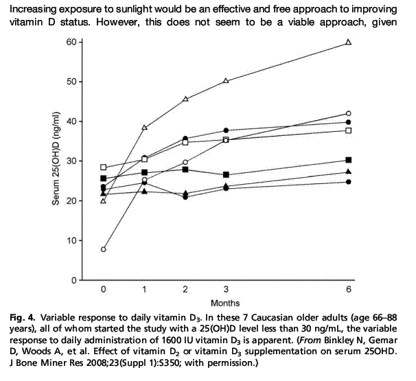
Low Vitamin D Status : Definition, Prevalence, Consequences, and Correction, Neil Binkley, Rekha Ramamurthy, Diane Krueger, pages 287-301, Abstract | Full Text | Full-Text PDF (292 KB) Vitamin D is obtained from cutaneous production when 7-dehydrocholesterol is converted to vitamin D3 (cholecalciferol) by ultraviolet B radiation or by oral intake of vitamin D2 (ergocalciferol) and D3. An individual's vitamin D status is best evaluated by measuring the circulating 25-hydroxyvitamin D (25(OH)D) concentration. Although controversy surrounds the definition of low vitamin D status, there is increasing agreement that the optimal circulating 25(OH)D level should be approximately 30 to 32 ng/mL or above. Using this definition, it has been estimated that approximately three-quarters of all adults in the United States have low levels. Low vitamin D status classically has skeletal consequences such as osteomalacia/rickets. More recently, associations between low vitamin D status and increased risk for various nonskeletal morbidities have been recognized; whether all of these associations are causally related to low vitamin D status remains to be determined. To achieve optimal vitamin D status, daily intakes of at least 1000 IU or more of vitamin D are required. The risk of toxicity with “high” amounts of vitamin D intake is low. Substantial between-individual variability exists in response to the same administered vitamin D dose. When to monitor 25(OH)D levels has received little attention. Supplementation with vitamin D3 may be preferable to vitamin D2. [tiki-download_file.php?fileId=1182]
Chart of variability of response to 1600 IU of vitamin D -click on thumbnail to see full size
Maternal Vitamin D Status : Implications for the Development of Infantile Nutritional Rickets, Kebashni Thandrayen, John M. Pettifor, pages 303-320, Abstract | Full Text | Full-Text PDF (526 KB) The mother is the major source of circulating 25-hydroxyvitamin D concentration in the young infant. Maternal vitamin D status is an important factor in determining the vitamin D status of the infant and their risk of developing vitamin D deficiency and infantile nutritional rickets. There is evidence that the current supplementation recommendations, particularly for pregnant and lactating women, are inadequate to ensure vitamin D sufficiency in these groups. A widespread and concerted effort is needed to ensure daily supplementation of breastfed and other infants at high risk with vitamin D 400 IU from birth and of pregnant women in high-risk communities with 2000 IU . Future studies are required to determine the optimal doses of vitamin D supplementation in pregnancy and during lactation, and for normalizing vitamin D stores in infancy to reduce the prevalence of infantile nutritional rickets. Operational research studies are needed to understand the best methods of implementing supplementation programs and the factors that are likely to impede their success.
Osteomalacia as a Result of Vitamin D Deficiency, Arti Bhan, Ajay D. Rao, D. Sudhaker Rao, pages 321-331, Osteomalacia is an end-stage bone disease of chronic and severe vitamin D or phosphate depletion of any cause. Its importance has increased because of the rising incidence of vitamin D deficiency. Yet, not all cases of osteomalacia are cured by vitamin D replacement, and furthermore, not all individuals with vitamin D deficiency develop osteomalacia. Although in the past osteomalacia was commonly caused by malabsorption, nutritional deficiency now is more common. In addition, recent literature suggests that nutritional vitamin D deficiency osteomalacia follows various bariatric surgeries for morbid obesity. Bone pain, tenderness, muscle weakness, and difficulty walking are all common clinical manifestations of osteomalacia. Diagnostic work-up involves biochemical assessment of vitamin D status and may also include a transiliac bone biopsy. Treatment is based on aggressive vitamin D repletion in most cases with follow-up biopsies if patients are started on antiresorptive or anabolic agents.
Common causes of vitamin D deficiency osteomalacia
Extrinsic Decreased exposure to sunlight, Use of sunscreens (especially >8 SPF), Use of a veil (or hijab), Increased or dark skin pigmentation, Inadequate dietary intake, Morbid obesity
Intrinsic Advancing age with decreased cutaneous production of vitamin D, Malabsorption caused by various gastrointestinal disorders, Gastrectomy (partial, total, or bypass procedure)
Acquired vitamin D deficiency (as a result of increased catabolism or metabolic clearance) [tiki-download_file.php?fileId=1168]
Genetic Disorders and Defects in Vitamin D Action, Peter J. Malloy, David Feldman , pages 333-346 Two rare genetic diseases can cause rickets in children. The critical enzyme to synthesize calcitriol from 25-hydroxyvitamin D, the circulating hormone precursor, is 25-hydroxyvitamin D-1?-hydroxylase (1?-hydroxylase). When this enzyme is defective and calcitriol can no longer be synthesized, the disease 1?-hydroxylase deficiency develops. The disease is also known as vitamin D–dependent rickets type 1 or pseudovitamin D deficiency rickets. When the VDR is defective, the disease hereditary vitamin D–resistant rickets, also known as vitamin D–dependent rickets type 2, develops. Both diseases are rare autosomal recessive disorders characterized by hypocalcemia, secondary hyperparathyroidism, and early onset severe rickets. In this article, these 2 genetic childhood diseases, which present similarly with hypocalcemia and rickets in infancy, are discussed and compared. [tiki-download_file.php?fileId=1171]
Vitamin D and Fracture Prevention , Heike A. Bischoff-Ferrari, pages 347-353, This article discusses the amount of vitamin D supplementation needed and the desirable 25-hydroxyvitamin D level to be achieved for optimal fracture prevention. [tiki-download_file.php?fileId=1173]
Vitamin D in Kidney Disease : Pathophysiology and the Utility of Treatment, Rizwan A. Qazi, Kevin J. Martin, pages 355-363, Vitamin D physiology has gained more importance and publicity than any of its counterparts in the water- and fat-soluble vitamin groups combined. This is partly because vitamin D deficiency is still widely prevalent in the developed world and the beneficial effects are thought to extend beyond the regulation of calcium and phosphorus homeostasis alone. Vitamin D deficiency becomes even more important in the various stages of chronic kidney disease (CKD); CKD itself is also on the increase. How vitamin D physiology is altered in CKD and how the various treatment modalities can alter the morbidity and mortality associated with CKD is the topic of discussion for this article. [tiki-download_file.php?fileId=1169]
Vitamin D and the Immune System : New Perspectives on an Old Theme, Martin Hewison, pages 365-379, Interaction with the immune system is one of the most well-established nonclassic effects of vitamin D. For many years this was considered to be a manifestation of granulomatous diseases such sarcoidosis, in which synthesis of active 1,25-dihydroxyvitamin D3 (1,25(OH)2D3) is known to be dysregulated. However, recent reports have supported a role for 1,25(OH)2D3 in mediating normal function of the innate and adaptive immune systems. Crucially, these effects seem to be mediated via localized autocrine or paracrine synthesis of 1,25(OH)2D3 from precursor 25-hydroxyvitamin D3, the main circulating metabolite of vitamin D. The ability of vitamin D to influence normal human immunity is highly dependent on the vitamin D status of individuals, and may lead to aberrant response to infection or autoimmunity in those who are lacking vitamin D. The potential health significance of this has been underlined by increasing awareness of impaired vitamin D status in populations across the globe. This article describes some of the recent developments with respect to vitamin D and the immune system, and possible clinical implications. [tiki-download_file.php?fileId=1166]
Vitamin D: Extraskeletal Health Michael F. Holick , pages 381-400. Vitamin D deficiency is the most common nutritional deficiency and likely the most common medical condition in the world. The major cause of vitamin D deficiency has been the lack of appreciation that the body requires 5- to 10-fold higher intakes than is currently recommended by health agencies . There is now overwhelming and compelling scientific and epidemiologic data suggesting that the human body requires a blood level of 25(OH)D above 30 ng/mL for maximum health. To increase the blood level to the minimum 30 ng/mL requires the ingestion of at least 1000 IU of vitamin D per day for adults . In general, there is no downside to increasing either a child's or adult's vitamin D intake. [tiki-download_file.php?fileId=1162]
The Role of Vitamin D in Cancer Prevention and Treatment , Aruna V. Krishnan, Donald L. Trump, Candace S. Johnson, David Feldman , Calcitriol (1,25-dihydroxyvitamin D3), the hormonally active form of vitamin D, exerts growth inhibitory and prodifferentiating effects on many malignant cells and retards tumor growth in animal models. Calcitriol is being evaluated as an anticancer agent in several human cancers. The mechanisms underlying the anticancer effects of calcitriol include inhibition of cell proliferation, stimulation of apoptosis, suppression of inflammation, and inhibition of tumor angiogenesis, invasion, and metastasis. This review discusses some of the molecular pathways mediating these anticancer actions of calcitriol and the preclinical data in cell culture and animal models. The clinical trials evaluating the use of calcitriol and its analogues in the treatment of patients with cancer are described. The reasons for the lack of impressive beneficial effects in clinical trials compared with the substantial efficacy seen in preclinical models are discussed. [tiki-download_file.php?fileId=1167]
Vitamin D and Diabetes , Tatiana Takiishi, Conny Gysemans, Roger Bouillon, Chantal Mathieu, pages 419-446, Type 1 (T1D) and type 2 (T2D) diabetes are considered multifactorial diseases in which both genetic predisposition and environmental factors participate in their development. Many cellular, preclinical, and observational studies support a role for vitamin D in the pathogenesis of both types of diabetes including: (1) T1D and T2D patients have a higher incidence of hypovitaminosis D; (2) pancreatic tissue (more specifically the insulin-producing ?-cells) as well as numerous cell types of the immune system express the vitamin D receptor (VDR) and vitamin D-binding protein (DBP); and (3) some allelic variations in genes involved in vitamin D metabolism and VDR are associated with glucose (in)tolerance, insulin secretion, and sensitivity, as well as inflammation. Moreover, pharmacologic doses of 1,25-dihydroxyvitamin D (1,25(OH)2D), the active form of vitamin D, prevent insulitis and T1D in nonobese diabetic (NOD) mice and other models of T1D, possibly by immune modulation as well as by direct effects on ?-cell function. In T2D, vitamin D supplementation can increase insulin sensitivity and decrease inflammation. This article reviews the role of vitamin D in the pathogenesis of T1D and T2D, focusing on the therapeutic potential for vitamin D in the prevention/intervention of T1D and T2D as well as its complications. [tiki-download_file.php?fileId=1165]
Vitamin D Analogs, Glenville Jones, pages 447-472, | Full-Text PDF (800 KB) Vitamin D has gone through a renaissance with the association of vitamin D deficiency with a wide array of common diseases including breast, colorectal and prostate cancers, cardio-vascular disease, autoimmune conditions and infections. Vitamin D analogs constitute a valuable group of compounds which can be used to regulate gene expression in functions as varied as calcium and phosphate homeostasis, as well as cell growth regulation and cell differentiation of a wide spectrum of cell types. This review will discuss the full range of vitamin D compounds currently available, some of their possible uses, and potential mechanisms of action.